Section: Surgical Oncology Curriculum: Curriculum, page 70
General
- Preferred treatment for many pts with end-stage renal disease
- Provides better quality of life than dialysis
- Releases pts from the dietary and fluid restrictions of dialysis and the physical constraints imposed by the need to dialyse
- More cost-effective and probably improves survival
- Frequently the primary cause of ESRF is unknown
Indications
(Causes of ESRF)
- Glomerulonephritis
- Diabetic nephropathy
- Hypertensive nephrosclerosis
- Renal vascular disease
- Polycystic disease
- Pyelonephritis
- Obstructive uropathy
- SLE
- Analgesic nephropathy
- Metabolic diseases
- Oxalosis, Amyloid
Pre-Transplant Evaluation
- All pts formally assessed by MDT team
- Incl. transplant surgeon and nephrologist
- Significant number are likely considered unsuitable because of major comorbidity, esp. cardiovascular disease
- Nature of the 1° renal disease does not generally affect the decision to proceed to transplantation
- No absolute upper age limit to transplantation
- However, older patients (over the age of 65 years) are less likely to be considered because of major comorbidity
- Cardiovascular assessment particularly important
- Cardiovascular disease very common in dialysis population
- Esp. those with diabetes
- The major cause of death after transplantation
- Cardiovascular disease very common in dialysis population
- Ensure urinary tract is functional and that there is no need for corrective urological surgery
- Only when there is intractable renal sepsis or very large polycystic kidneys that intrude into both iliac fossae is native nephrectomy required before transplantation
- Make sure able to cope psychologically with transplantation and likely to comply with immunosuppressive therapy
- Contraindications:
- Pre-existing malignancy (wait 3yr till after treatment)
- Active infection
Technique of renal transplant
- Placed in the iliac fossa in the experitoneal position
- Leaving the native kidneys in-situ
- Vascular
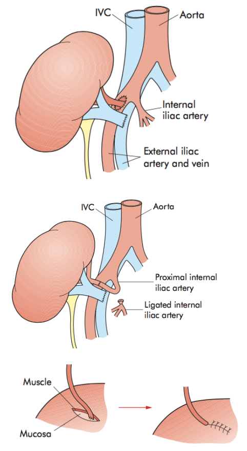
- Donor Renal Vein is anastomosed end-to-side to the External Iliac Vein
- If from deceased donor, Renal Artery will come with Carrel Patch
- Patch of Aorta that gives rise to Renal Artery/arteries
- Anastomosed end-to-side to the External Iliac Artery
- If the donor renal artery lacks an aortic patch, (living donor transplant)
- Anastomose the donor artery end-to-end to the recipient Internal Iliac Artery
- Following completion of the venous and arterial anastomoses, the vascular clamps are removed and the kidney is allowed to reperfuse with blood
- Ureter
- Kept reasonably short to avoid the risk of distal ischaemia
- Direct implantation of the ureter into the dome of the bladder
- Mucosa-to-mucosa anastomosis
- Followed by closure of the muscular wall of the bladder over the ureter to create a short tunnel
- JJ ureteric stent left in-situ
- Reduce the risk of urine leak or early obstruction
- Before closing, important to check is in satisfactory position
- Without kinking/torsion
Technical Complications
Vascular
- Incidence of vascular complications low
- Renal Artery Thrombosis (1%)
- Renal Vein Thrombosis is more common (up to 5% of cases)
- Aetiology often uncertain
- Sometimes from technical error
- Presents during the 1st week
- Sudden pain and swelling at the site of the graft
- Diagnosis confirmed by Doppler USS
- Urgent surgical exploration indicated
- In most cases, transplant nephrectomy is required
- Can be minimised by giving low-dose heparin or aspirin prophylaxis
- Aetiology often uncertain
- Renal Artery Stenosis (up to 10%)
- Usually presents late (often years)
- Get increasing hypertension and decreasing renal function
- Diagnosed by angiography
- Best treated by angioplasty
- If unsuccessful or not technically possible, may need open surgery and vascular reconstruction
Urological
- Urological complications ~ 5% in the early post-transplant period
- Incidence can be reduced markedly by leaving temporary ureteric stent
- Urinary leaks
- Result from technical errors at the ureteric anastomosis or ureteric ischaemia
- Present with discomfort and leakage of urine from the wound
- Usually require surgical intervention
- Reimplantation of the ureter into the bladder
- Or anastomosis of the transplant ureter to the ipsilateral native ureter
- Obstruction of the transplant ureter
- May occur early or late
- Causes:
- Technical error
- External pressure from a haematoma or lymphocele
- Ischaemic stricture
- Presents with painless deterioration of graft function
- Diagnosed on USS
- Hydronephrosis and ureteric dilatation
- Initial treatment is by percutaneous antegrade nephrostomy
- And sometimes insertion of a stent
- Some amenable to balloon dilatation
- Most are best treated by surgical intervention
Lymphocoele
- Usually asymptomatic
- Occasionally large enough to cause ureteric obstruction or oedema of the ipsilateral leg
- Initial treatment
- Ultrasound-guided percutaneous drainage
- In large or recurrent lymphoceles
- Repeated instillation of povidone-iodine into the lymphocele cavity by a percutaneous drain
- Surgical intervention may be needed to drain a persistent lymphocele into the peritoneal cavity
Renal Transplant Consent
- Complications
- Whitebook, page 122
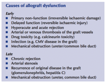
Causes of Graft Dysfunction
Investigation of Graft Dysfunction
- Delayed Graft Function
- Definition: Need for dialysis post- transplantation as a result of acute tubular necrosis
- Incidence:
- 30% of heart-beating deceased donors
- Up to 50% of NHBD kidney transplants
- Uncommon (< 5%) following living donor transplantation
- Oliguria may be useless indicator as recipients may produce significant urine from their native kidneys
- Delayed function minimised by:
- Optimizing donor management before kidney procurement
- Reducing the cold ischaemia time
- Management of Early Graft Dysfunction
- First step - urinary catheter should be irrigated in case it is occluded by a blood clot
- Hypovolaemia fixed and CVP monitors
- Calcineurin inhibitors often withheld/reduced
- Investigations
- Doppler USS of graft is single most important investigation
- Allows exclusion of vascular thrombus and urinary obstruction as causes
- Radionucleotide scan may give information of perfusion and excretion
- If dysfunction still present after a few days
- USS-guided biopsy
- ATN usually resolves within 4 weeks
- < 5% suffer primary non-function and never regain function
- Doppler USS of graft is single most important investigation
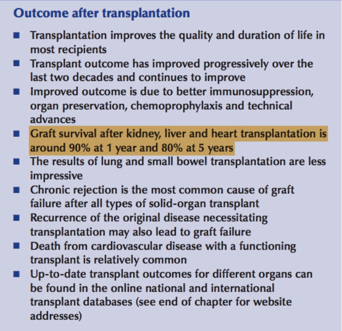
Outcome After Transplant