-
Sleeve technique
-
or Dorsal slit
-
Local anesthesia and skin asepsis are performed
-
Marking
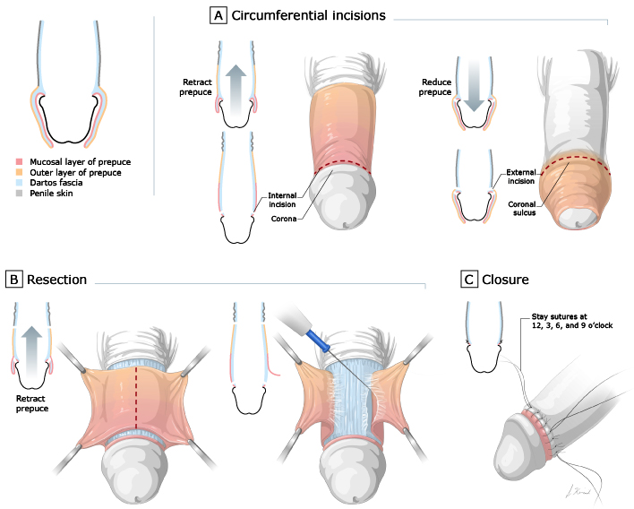
- Two incision sites are marked circumferentially
- One on the mucosal side of the fοrеskiո with the рrерսсe in a retracted position, leaving a 5 to 10 mm cuff around the base of the corona, and following the natural V shape of the frenulum on the ventral side.
- Second skin marking is then made on the outer aspect of the fοrеѕkiո around the base of the corona while it is reduced back to its normal position. It is important not to remove too much preputial skin, as penile shortening and tethering may occur postoperatively.
- Two incision sites are marked circumferentially
-
Incision
- Both incisions are made with a #15 scalpel blade and carried through the Dartos fascia
- The fοrеskin is then incised in a longitudinal fashion, adjoining both circumferential incisions, and is resected circumferentially using either scissors or electrocautery. The fοreskin is usually sent to pathology for formal analysis, especially if there is underlying preputial disease.
-
Hemostasis is performed using monopolar or bipolar electrocautery.
-
The skin edges are then approximated using interrupted 4-0 or 5-0 resorbable sutures