Section: Vascular Sub-section: Curriculum, page 100
Overview
- Aneurysm Definition = 50% dilated compared to adjacent vessel
- A true aneurysm involves primary dilatation of the artery including all vessel wall layers (intima, media & adventitia)
- False aneurysm / pseudoaneurysm = Disruption of the artery wall (Really a walled-off haematoma)
Aetiology
- Congenital
- Weak areas – Berry aneurysm
- Weak materials – Ehlers-Danlos Syndrome, Marfan Syndrome
- Acquired
- Traumatic
- Infection (mycotic) – e.g. Syphilis, Staphylococcus, Salmonella
- Metabolic
- Pregnancy
- Spleen, liver, renal, aorta
- Usually presents with shock, MR 65%
- Pregnancy
- 90% Degeneration
- Atherosclerosis – 90% of aneurysms - but relationship foggy
- Age – Elastin half-life 40-70 years – not re-synthesized
- Cystic Medial Necrosis
Classification
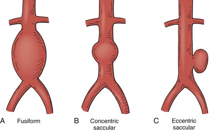 - Fusiform
- Uniform in shape with symmetrical dilatation that involves the entire circumference of the aortic wall
- Fusiform
- Uniform in shape with symmetrical dilatation that involves the entire circumference of the aortic wall
- Saccular
- Localized and appears as an outpouching of only a portion of the aortic wall.
- Saccular aneurysms may be a manifestation of plaque hemorrhage and aortic ulceration or aortic wall infection and appear to have an increased risk for rupture
- Localized and appears as an outpouching of only a portion of the aortic wall.
Pathology
- Atherosclerosis
- Destruction of Elastin in media
- Collagen will then hold its integrity but when the collagen goes…RUPTURE
- Macrophages secrete Metalloproteinases
- These destroy Elastin and collagen
- Results in gradual dilation of vessel wall
- Further endothelial damage due to increased wall tension and increased turbulent flow
- Cascade of damage and dilation
- Laplace’s law: Transmural pressure (P) = Tension / radius
- RUPTURE
- Destruction of Elastin in media
- Hypertension
- Causes luminal narrowing of the vasa vasorum that supply artery
- Leads to ischeamia of the outer layers of the media
- Leads to degeneration and fibrosis
- External injury
- Mycotic aneurysms - septic embolis
- Trauma
- Vasculitis - antibody mediated injury
- Areas
- Infrarenal aorta has a lower concentration of lamellar units of elastin & collagen than other areas
- Morphology – saccular versus fusiform
- AORTA > ILIAC > POPLITEAL > FEMORAL > THORACIC > CAROTID > SUBCLAVIAN > SPLANCHNIC (Spleen, Liver, Renal) > CEREBRAL
Aortic Aneurysm
- Definition: Aneurysmal dilatation of the Abdominal Aorta (2.6cm female, 3cm male)
- Aorta does increase in size with age & weight
Incidence
- Men 65-80 - 3-5%
- < 60yo = Negligible
- M:F ratio 5:1.
- 5-10% Men, 3% women
- 50% of pts with femoral / popliteal aneurysms also have AAA
Classification
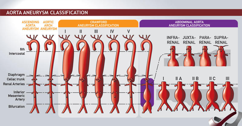
Thoracic
- Ascending aortic aneurysms
- Arise anywhere from the aortic valve to the brachiocephalic trunk (ie, innominate artery; 60 percent)
- Aortic arch aneurysms
- Include any thoracic аոeuryѕm that involves the brachiocephalic vessels (10 percent)
- Descending aortic aneurysms
- Distal to the left subclavian artery (40 percent)
- Thoracoabdominal aneurysms (10 percent)
Thoraco-abdominal Aneurysm
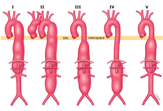
The Crawford classification of TAAA is based upon the extent of aortic involvement.
- Type I
- Arises from above the sixth intercostal space
- Extends to include the origins of the celiac axis and superior mesenteric arteries.
- Does not extend into the infrarenal aortic segment.
- Type II aneurysm
- Arises above the sixth intercostal space
- May include the ascending aorta
- Extends distal to include the infrarenal aortic segment
- Type III aneurysm
- Arises below the sixth intercostal space
- Extends into the abdominal aorta.
- Type IV aneurysm
- Involves the entire abdominal aorta from the level of the diaphragm to the aortic bifurcation.
- Type V aneurysm
- Arises below the sixth intercostal space
- Extends into the abdominal aorta, but is limited to the visceral segment.
Abdominal (90%)
- Classification
- Infrarenal
- Originates below the renal arteries
- Juxtarenal
- Originates at the level of the renal arteries, but the aorta at the renal arteries is normal.
- Pararenal
- Involves the aorta at the level of the renal arteries (ie, the renal artery originates from an aneurysmal aorta
- Suprarenal (visceral)
- Aոeսryѕm originates above the renal arteries.
- Infrarenal
- Iliac vessels involved in 40%.
- 25% co-existing fem/pop aneurysm
Inflammatory
- 5% of all AAA
- Thickened aneurysm wall and peri-aneurysmal fibrosis with dense visceral adhesions
- Often involves D4, IVC, Left Renal Vein
- Aetiology unknown – associated with autoimmune conditions, raised ESR
Aetiology
- Risk Factors
- Age
- M > F 5:1
- Smoking (AAA preponderance – 8:1 smokers vs non-smokers)
- Family hx: +ve ⇒ 20% chance 1° relative has AAA
- Ethnic origin
- Elastin/collagen abnormality: Marfans, Ehlers-Danlos
- Hypertension: small effect; 40% with AAA
- Atherosclerosis
- NB: NOT DM – lower incidence of AAA
- Other Causes
- Mycotic
- Cystic medial necrosis
- Arteritis
- Trauma
- Inherited CT disorders
Clinical
- Most asymptomatic. 75% die before admission to hospital
- Many detected incidentally
- Rupture – 70% that present did not know they had an aneurysm
- Triad
- Pain (abdo, back, groin)
- Collapse / syncope / hypotension
- Pulsatile (tender) mass
- Severe pain in the absence of rupture characterizes the rare inflammatory aneurysm that is surrounded by 2-4cm perianeurysmal retroperitoneal inflammatory reaction
- Inflammation usually confined to the anterior aorta & iliacs
- Seen on CT as a characteristic thick wall
- Endovascular repair is the procedure of choice for inflammatory aneurysms
- Severe tenderness suggests rupture / inflammation / recent expansion → Urgent imaging & surgery usually required
- Triad
- Thromboembolic complications are uncommon (2-5% of presentations)
- Thrombi in Aorta aneurysms rarely embolise
Investigation
- Incidental finding
- On Plain X-ray: Eggs-shell calcification in the outer layers of ≈ 2 0% of AAAs
- On USS. USS good for screening.
- CT-A = Investigation of choice
- Evaluation for endoluminal repair
- Spiral CTA with 3D reconstruction
- Extent of disease
- Calcification
- Presence of dissection
- Identifies occlusive disease
- Accessory renal vessels
- Presence of retroperitoneal fibrosis
- Relationship to surrounding structures
- Extravasation
- Evaluation for endoluminal repair
Features suitable for endovascular repair:
- Favourable anatomy
- This is steadily expanding with improvements in techniques
- Higher level of perioperative risk
- Most benefit is in the short term
- Open sսrgiϲal repair may be preferred for younger patients who have a low or average perioperative risk.
Screening
- No official screening programme in NZ
- Screening program in the UK based of the The Multicentre Aneurysm Screening Study (MASS) funded by the UK Medical Research Council
- Screening decreases aneurysm related death
- Hazard ratio 0.49 vs 0.85 per 1000 person years
- @ 4yrs: 40% risk reduction (Deaths = 0.19% vs 0.33%)
- No benefit by 15yrs of screening
- Main issue = Cost
- NNT (to save one life) ≈ 710
Risk of Rupture
- < 5cm = <1% rupture/year
- 5 - 5.9 = 3%
- 6 - 6.9 = 10%
-
7cm = 25-30%
Management
- Acute AAA:
- IV access, X-match, IDC
- “Hypotensive haemostasis”: Aim for consciousness / SBP > 80mmHg
- Boluses of crystalloid
- Laparotomy for ruptured AAA, especially if haemodynamically unstable
- Endovascular аոeurуѕm repair reduces perioperative (30 day) morbidity and mortality following elective AAΑ repair and there is accumulating evidence that morbidity and mortality following repair of symptomatic or ruptured AAΑ may also be reduced
- Contained leak (retroperitoneal rupture) may tamponade itself for 6-8hrs Median survival without Rx ≈ 8hrs
- Elective
- Indications
- Asymptomatic AAA ≥5.5 cm in maleѕ; asymptomatic AAA ≥5.0 in femаlеѕ>
- No evidence to repair small aneurysms 4-5.5cm
- UK Small Aneurysm Trial showed that early surgery for aneurysms < 5.5cm concurred no long-term survival benefit over regular surveillance
- Rapid expansion (> 0.5cm / 6 months)
- Symptomatic – regardless of size
- Asymptomatic AAA ≥5.5 cm in maleѕ; asymptomatic AAA ≥5.0 in femаlеѕ>
- Contraindications
- Life expectancy < 2 years
- Recent MI, intractable Heart Failure/ Angina
- Severe pulmonary insufficiency
- Severe CRF
- Incapacitating Stroke
- Behçet’s (Genital, mucosal ulceration & vasculitis – suture lines break down)
- Life expectancy < 2 years
- Indications
Miscellaneous
- If pt has co-existent Bowel Ca → Weigh up risk of AAA rupture vs risk of colonic obstruction
- Consider endoluminal Rx
- NB: AAA likely to ‘soften’ due to operative inflammation from bowel surgery (therefore risk ↑ peri-op)
- If pt has active IHD
- Investigate & treat prior to elective AAA repair
- IMA often occluded
- Colonic ischemia is uncommon
- The adequacy of perfusion to the left colon should always be assessed prior to completion of the case
- The inferior mesenteric artery should be reimplanted if there is any question of bowel viability.
- In patients with prior colon resection or reduced inflow to one or both internal iliac arteries, reimplantation of the inferior mesenteric artery is appropriate.
Endovascular Repair (EVAR)
- Fewer short-term complications (incl. immediate mortality) 1% vs 5%
- More long-term (↑ risk of rupture & re-intervention rates)
- Reintervention highest in 1st 6/12
- Delayed rupture in 2% vs 0% in open
- Long-term complications after open repair are not usually related to the aneurysm – e.g. hernias, SBO
- Overall survival: no diff at 4yrs
EVAR-1 trial (UK Lancet 2005):
- Endovascular vs open repair, ≥ 5.5cm
- ↓ immediate complications & 30-day mortality in endovascular (1.5 vs 4.6%)
- ↑ post-operative complications & re-intervention in endovascular group (40 vs 10%)
- But no difference in overall survival (50% survival in both groups at 8yrs)
Requirements for endovascular repair
- CT angio – to measure lengths/diameters to select appropriate size endograft
- Suitable proximal & distal attachments sites
- Adequate access to arteries
Exclusion criteria for EVAR
- Proximal neck
- < 15mm long or > 30mm diameter or > 60° angulation
- Iliac artery fixation site
- < 10mm long, iliac tortuosity ++ or iliac artery < 8mm or > 24mm in diameter
- Indispensable IMA
- Inability to maintain at least one patent internal iliac artery
Complications
- Site
- Endoleak
- Migration → Leak
- Fenestrated devices (F-EVAR) available for juxtarenal (= Short < 10mm or no infrarenal neck) AAAs
Post-EVAR Surveillance
- Lifelong Surveillance – CT at 1 month then USS/AXR at 3-6 months for 2 yrs then annually for life
Prognosis
- Aneurysms increase in size by 0.5cm per annum
- Higher expansion rate in:
- Larger aneurysms
- Women x 2-3
- Increased age
- Smokers 13-20%
- COPD
- Hypertension (Rate of expansion correlates with diastolic BP)
- Higher expansion rate in:
Risk of Rupture Relates to
Size
| Size (cm) | 1yr rate of rupture (%) | 5-yr rate of rupture (%) |
|---|---|---|
| <5 5-6 6 7+ | ≈<1% 5% 10% 30% | 10 % 25 % 35 % 75% |
| Expansion |
-
0.5cm over 6 months
Elective
- MR 1-5%
- 30-Day Significant Complication rate 10%
- ↑ Mortality for repair of Juxtarenal aneurysms (2.5-6%)
- Endovascular
- MR 2% (30-Days)
- 6-Month Complication Risk: 40%
- Emergency
- MR 40% of those that make it to surgery
- 75% die before hospital
- Prognostic Factors
- pH < 6.8
- Age > 75
- Creatinine > 132 (independent RF for 20 day MR (5.5%) for endovascular & open repair)
- Hb <90
- LOC
- ECG ischemic changes
- Suprarenal Aneurysms
- Operative MR 2 x Infrarenal aneurysms
- Renal failure & bowel ischaemia more common
- Can use fenestrated grafts or anastomose vessels to graft to enable flow
- Scoring system
- No perfect scoring system for predicting mortality, includes POSSUM & its variations
- Glasgow Aneurysm Score: Predicting mortality after AAA repair
- GAS = (Age in years) + (17 for shock) + (7 for myocardial disease) + (10 for cerebrovascular disease) + (14 for renal disease)
- Score > 85 gives high chance of mortality
Endoleak
- Persistent blood flow outside the lumen of the endoluminal graft but within an aneurysm sac or adjacent vascular segment being treated by the device
Aetiology
- Due to incomplete sealing, or exclusion of the aneurysm sac, and thus cause reflux of blood flow into the sac
Classification
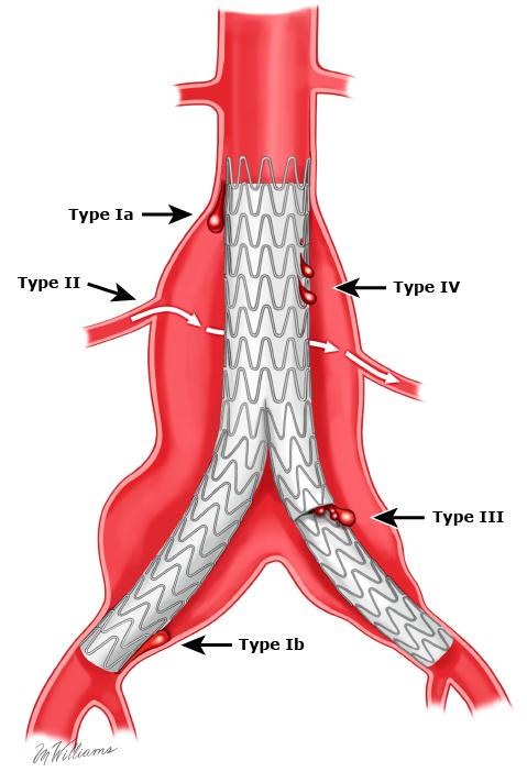
- Type I- Incompetent seal
- At the proximal (Ia) or distal (Ib) attachment site.
- Usually occurs in early course of treatment, but may occur later, due to stent/graft migration
- Should be fixed immediately
- At the proximal (Ia) or distal (Ib) attachment site.
- Type II results - Patent branch vessels
- From flow into and out of the aneurysm sac from one or more artery (lumbar or inferior mesenteric artery)
- In some circumstance - 2 or more patent vessels present - inflow and outflow develops - active blood flow within channel within the aneurysm sac
- Unless enlarging, can be treated conservatively
- Rx: Coil embolization, N-butyl cyanoacrylate glue, Retroperitoneal endoscopic ligation or Open approach
- From flow into and out of the aneurysm sac from one or more artery (lumbar or inferior mesenteric artery)
- Type III - Dissociation of modular components
- Inadequate / ineffective sealing of overlapping graft joints or rupture of the graft fabric
- Occurs early due to technical problems, or later due to device breakdown
- If enlarging, the graft should be replaced
- Type IV - Porous graft material.
- Causing blood to pass through from graft to sac
- Endoleak of undefined origin (type V) is continued aneurysm sac expansion without a demonstrable endoleak on any imaging modality. It is also referred to as endotension.
- Requires an angiogram / CT-Angio to accurately diagnose & determine type
Thoracic Aortic Aneurysm
- Dilatation of thoracic aorta 1.5x
Incidence
- M > F
- ≈ 3% of pts > 65yo
Classification
- Ascending aortic aneurysms
- Arise anywhere from the aortic valve to the brachiocephalic trunk (ie, innominate artery; 60 percent)
- Aortic arch aneurysms
- Include any thoracic аոeuryѕm that involves the brachiocephalic vessels (10 percent)
- Descending aortic aneurysms
- Distal to the left subclavian artery (40 percent)
- Thoracoabdominal aneurysms (10 percent)
Aetiology
- Arteriosclerosis
- Medial degeneration of the aortic wall
- Marfans syndrome
- Trauma
- Infection
Clinical
- Usually have a history of HTN
Pathology
- Medial degeneration of the aortic wall: Replacement of muscle cells & elastic lamina with mucoid-filled cystic spaces
Investigations
- CXR: Convexity at R heart border
- CT
- MRA
- Coronary angiogram – to screen for coexistent coronary artery disease
Management
- Asymptomatic patients
- Sporadic
- If <5.5 Aggressive BP control and surveillance
- If Aneurysm > 5.5 cm & asymptomatic = consider surgery
- Marfan’s Syndrome
- Intervention recommended at 4.5-5cm depending on risk factors
- Other risk factors to consider small size of intervension
- аոeսrуѕm expansion >0.5 cm per year, saccular аոеurуѕm, infectious аոеսrуѕm, female sex
- Approach
- Ascending Aorta
- Open surgery using cardiopulmonary bypass and sometimes requires aortic root replacement with coronary artery reimplantation]
- Descending Aorta
- EVAR or Open
- Ascending Aorta
Prognosis
- If untreated ≈ 50% of non-dissecting thoracic aneurysms will rupture
- ↑ Risk of rupture:
- Size
- Underlying CT disorder
- Dissection
- COPD
- Advancing age
Iliac Aneurysm
Incidence
- In conjunction with AAA (Isolated iliac aneurysms unusual – usually CIA/IIA not EIA)
Clinical
- Most are asymptomatic & incidental
- May present with symptoms related to compression / erosion of surrounding structures
- Obstructive uropathy - ureteral obstruction / neuropathy / unilateral leg swelling - adjacent iliac vein
- Symptomatic aneurysms / > 4cm may be palpable on abdominal or rectal exam
Management
- Diameter ≥3 cm→ Repair if pt fit
- Repair aims to preserve flow to pelvis through at least one Internal Iliac Artery to prevent buttock claudication / impotence and maintain perfusion to the distal colon
- Open vs endovascular repair
- Endovascular repair preferred
- Open repair of isolated iliac arteries can be done though a retroperitoneal approach
Popliteal Aneurysm
- Increase in diameter 1.5 x = > 15mm
Incidence
- Account for 80% of peripheral arterial aneurysms
-
90% Male
- Mean age 65yrs
- Overall prevalence < 1%
- 50% bilateral and 40% associated with AAA
Aetiology
- Atherosclerosis (most)
- Trauma
- Familial
Clinical
- 50% symptomatic (thrombosis, embolism, compression, rupture)
- Pts present with acute (20%) or chronic (15%) limb ischaemia secondary to aneurysm thrombosis or distal emboli
- Rupture is rare
- Often have had long hx of microemboli resulting in significant downstream damage – thus, often poor run-off vessels
- Symptoms may be transient
- Clinically: Easily palpable popliteal pulse
Investigations
- Duplex USS/ CT
- Angio: May not demonstrate the aneurysm well due to mural thrombus reducing lumen diameter
Management
- In acute ischaemia
- Emergency popliteal аnеսrysm repair
- intra-arterial thrombolysis
- If absent or limited distal runoff
- Restoration of outflow improves the long-term patency of revascularization
- Τhrοmbоlytiϲ agents can be administered intraoperatively or using catheter-based techniques
- Can prepare pt for surgery or be done when immediate surgery is not imperative (i.e. light touch intact) – good to clear run-off vessels in-order to bypass graft
- Indications for surgical reconstruction (ligation and vein bypass)
- 2cm
- Presence of laminar thrombus
- If symptomatic with peripheral embolization
- Approach
- Posterior approach with inlay autologous graft is most preferred
- Bypass graft and tie off often results in ongoing flow into aneurysm and dilation of aneurysm
- Endovascular repair is a viable option now given flexible endografts for symptomatic pts in the elective setting – though may have risk of disruption of thrombus and downstream emboli
Prognosis
- High complication rates (60%) if left untreated
- Majority will become symptomatic within 1-3yrs if left untreated
- Pts who develop acute ischaemia have a relatively poor prognosis (15% amputation rate)
- 5yr graft patency rates after surgical repair for asymptomatic pts ≈ 80%
Follow-up
- Lifelong, careful surveillance post-op or any asymptomatic pt with aneurysm
- Includes to detect and treat new aneurysms at other sites
Subclavian artery aneurysm
- Causes
- Trauma
- Thoracic outlet syndrome
- instrumentation
- Connective tissue disorders
- Iոfеction (ie, mycotic аոeurуѕm),
- Fibromuscular dysplasia
- Tumor
- Vasculitis
- Trauma
- Clinical
- Most subclavian artery aneurysms are asymptomatic, presenting as a pulsatile supraclavicular mass
- Symptoms occur
- Ischemic manifestations of distal embolization.
- Macroemboli can cause acute or chronic upper extremity ischemia
- Microemboli can cause digital symptoms including subungual splinter hemorrhages or digital pain, discoloration, or ulceration.
- Ischemic manifestations of distal embolization.
- Managemnt
- Symptomatic
- Repair
- Asymptomatic
- If intraluminal thrombus should be repaired
- Small asymptomatic aneurysms without intraluminal thrombus may be managed conservatively and evaluated with serial imaging - no consensus on size however > x2 increase in size is used
- Options
- Open or endovascular
- Symptomatic
Kommerell’s diverticulum
- Aneurysmal degeneration of the origin of an aberrant subclavian
- Arteria Lusoria)
- Incidence 0.5%
- Arises from Aorta distal to Left Subclavian Artery and courses behind the oesophagus
- Associated with a non-recurrent laryngeal nerve
- May compress the Oesophagus against the Trachea → “Dysphagia Lusoria”
- Arteria Lusoria)
- Clinical
- Typically asymptomatic
- Symptoms
- Dуѕрhаgia from esophageal compression by the аոеuryѕm (termed ‘ԁyѕрhagia lusoria’)
- Cough or ѕtridоr from tracheal irritation or compression
- Chest pain due to аոeսrуsm expansion or rupture
- Symptoms of ischemia such as exercise-induced аrm fatigue
- Treatment
- Almost always Repair - resection of the restricting structure & arterial replacement
- No guideline regarding the diameter for asymptomatic Kommerell diverticulum given its rarity, but a diameter threshold of >3 cm has been proposed
- Options
- Open or endovascular
- Almost always Repair - resection of the restricting structure & arterial replacement
Axillary artery aneurysm
- Incidence and Causes:
- Axillary artery aneurysms are rare, most often associated with traumatic arterial injuries (acute or chronic). Other causes include atherosclerosis, arteritis, connective tissue disorders, congenital factors, or idiopathic etiologies.
- Presentation:
- Often asymptomatic, presenting as a pulsatile axillary mass.
- Symptoms may include arm pain or dysfunction due to distal embolization or nerve compression.
- Rupture is rare but possible.
- Diagnosis:
- Physical Exam
- Palpation of the infraclavicular or axillary region to detect masses; evaluation of the arm and hand for distal embolization signs.
- Imaging
- Ultrasound is the primary diagnostic tool. CT or MR angiography aids in diagnosis and surgical planning. Digital subtraction angiography (DSA) is used selectively for evaluating distal arterial runoff.
- Physical Exam
- Management
- Indications for Repair:
- Symptomatic aneurysms or Asymptomatic aneurysms that are twice the diameter of the normal artery.
- Surgical Repair:
- Open surgical repair with interposition grafting.
- Preferred approach:
- Care is required due to proximity to the brachial plexus.
- Conduits:
- Prosthetic for large arteries and short repairs.
- Autogenous vein grafts for small arteries and long repairs.
- Open surgical repair with interposition grafting.
- Endovascular Repair:
- Less common due to motion and compression of the axillary artery.
- Reserved for focal aneurysms, palliation, or bridging in acute settings.
- Covered self-expanding stents are used to avoid external compression.
- Indications for Repair:
Brachial artery aneurysm
- Incidence
- Extremely rare, primarily caused by trauma.
- Cause
- Commonly associated with pseudoaneurysms due to:
- Dialysis access (current or former arteriovenous fistulas).
- Illicit drug injections.
- Other causes: connective tissue disorders, atherosclerosis, infection, congenital factors, or idiopathic origins.
- Commonly associated with pseudoaneurysms due to:
- Presentation:
- Typical finding: Pulsatile mass, either asymptomatic or symptomatic.
- Symptoms:
- Pain, numbness, paresthesias.
- Sensorimotor deficits in the median nerve distribution (digits 1–3).
- Ischemic symptoms from peripheral emboli (forearm, hand, fingers).
- Rupture: Rare but possible, particularly in pseudoaneurysms from drug injection.
- Diagnosis:
- Physical Exam: Pulsatile mass in the medial upper arm; assess for distal ischemia (pulse examination, signs of embolization, nerve compression).
- Imaging:
- Duplex ultrasound for initial evaluation.
- CT/MR angiography for thrombus identification and inflow/outflow assessment.
- Digital subtraction angiography for operative planning, particularly to evaluate distal runoff.
- Management
- Indications for Repair:
- Symptomatic aneurysms.
- Asymptomatic aneurysms twice the diameter of the adjacent artery.
- Indications for Repair:
- Surgical Repair:
- Preferred approach: Open surgical repair.
- Techniques:
- Focal aneurysms: Resection with primary repair.
- Most cases: Interposition bypass graft, usually with an autogenous conduit (preferred due to durability) or prosthetic conduit if necessary.
- Focal aneurysms: Resection with primary repair.
- Endovascular Repair:
- Limited use due to risks of stent fractures (superficial location and frequent artery flexion).
- Best suited for pseudoaneurysms, particularly at dialysis access sites.
- Durability is less favorable compared to surgical repair.
Innominate Artery Aneurysms
- Incidence and Causes:
- Rare, accounting for 2–5% of all aortic arch vessel aneurysms and <1% of all peripheral artery aneurysms.
- Most commonly caused by degenerative disease/atherosclerosis (50% of cases).
- Other causes: syphilis, tuberculosis, Kawasaki disease, Takayasu arteritis, Behcet disease, connective tissue disorders (e.g., Marfan, Ehlers-Danlos), trauma (pseudoaneurysms), and infection (mycotic aneurysms).
- Presentation:
- Often discovered incidentally; 25% present with symptoms.
- Symptoms:
- Pain (from expansion or rupture).
- Ischemic symptoms: transient ischemic attack (TIA), stroke, limb ischemia, or tissue loss.
- Compression symptoms: brachial plexus (pain, dysfunction), right recurrent laryngeal nerve (hoarseness), trachea (dyspnea), cervical sympathetic chain (Horner syndrome), or lung apex (hemoptysis).
- Rupture: Rare (10%), but life-threatening.
- Diagnosis:
- Physical Exam
- Supraclavicular bruit, absent pulses, signs of distal embolization, neurologic deficits, or compression symptoms.
- Imaging
- CT angiography is the gold standard for evaluating size, thrombus, and anatomical relationships.
- Physical Exam
- Classification:
- Type A: Involves only the innominate artery.
- Type B: Involves the innominate artery and its origin.
- Type C: Involves the innominate artery, its origin, and the ascending aorta (requires cardiopulmonary bypass).
- Management :
- Indications for Repair:
- Symptomatic aneurysms.
- Asymptomatic saccular aneurysms or fusiform aneurysms >3 cm in diameter.
- Surgical Repair:
- Open repair is the standard but involves significant morbidity due to the need for a Median sternotomy or cardiopulmonary bypass.
- Conduits: Prosthetic grafts (PTFE or Dacron) are preferred, except for mycotic aneurysms where autogenous grafts are used.
- Open repair is the standard but involves significant morbidity due to the need for a Median sternotomy or cardiopulmonary bypass.
- Endovascular Repair:
- Less invasive but limited by anatomical constraints (proximal/distal seal zones).
- Risks: Compression and fracture due to the mobile nature of the thoracic outlet.
- Stents: Balloon-expandable or self-expanding covered stents may be used selectively.
- Indications for Repair:
Carotid artery aneurysm
- Incidence
- Rare, accounting for 0.4–4% of aortic arch vessel aneurysms.
- Causes
- Degenerative disease/atherosclerosis (most common, often with hypertension), trauma (blunt, penetrating, or iatrogenic), arteritides (e.g., Takayasu arteritis, Behcet disease), connective tissue disorders (e.g., Marfan, Ehlers-Danlos), radiation injury, fibromuscular dysplasia, and infection.
- Presentation:
- More likely symptomatic compared to other arch vessel aneurysms (14–43% symptomatic).
- Symptoms:
- Pain (expansion or rupture).
- Cranial nerve palsy or Horner syndrome from compression.
- Dyspnea from tracheal compression.
- Neurologic deficits (amaurosis fugax, TIA, stroke) from thromboembolism.
- Proximal aneurysms are too deep to palpate; distal ones may be palpable or associated with a bruit.
- Diagnosis:
- CT angiography is the preferred diagnostic method, providing details on aneurysm size, thrombus burden, and anatomical relationships.
- MR angiography is an alternative, and any ultrasound-identified aneurysm should be further evaluated with advanced imaging.
- Management :
- Indications for Repair:
- All common carotid artery aneurysms should be repaired due to the high risk of embolic stroke.
- Nonoperative observation is associated with unacceptable morbidity and mortality.
- Open Surgical Repair
- Standard treatment, requiring exposure via median sternotomy.
- Preferred graft: Prosthetic (PTFE) for proximal aneurysms; autogenous vein graft for distal aneurysms or those involving the bifurcation.
- Standard treatment, requiring exposure via median sternotomy.
- Endovascular Repair:
- Less invasive, avoiding sternotomy, but requires favorable anatomy (adequate proximal and distal seal zones).
- Covered nitinol stents (balloon-expandable or self-expanding) are used selectively.
- Clinical outcomes from open, endovascular, and hybrid approaches are generally good, as demonstrated in meta-analyses
- Indications for Repair:
Pseudoaneurysm / False Aneurysm
- Definition
- Disruption of the artery wall = Pulsatile haematoma not contained by the artery wall but by a fibrous capsule
- Incidence
- Occurs in 1% after femoral catheterization
- Aetiology
- Follows disruption of a vessel: e.g. post-op or following a puncture / stab wound
- Clinical
- Thrombosis and embolization are the main risk
- Can cause venous compression + oedema/DVT
- Rupture is rare
- Pathology
- Haematoma forms outside the artery and thrombus initially plugs the hole in the artery but pulsatile blood pressure gradually forces the haemostatic plug outwards into the hematoma to form a cavity connected to the lumen of the vessel
- Investigations
- Duplex USS
- CTA
- Management
- Pseudoaneurysm < 3cm likely to spontaneously resolve
- After arterial puncture < 3cm usually resolve within 4 weeks; require USS f/up
- Larger aneurysms → Operative repair or Endovascular Embolization
- Acute pseudoaneurysm after femoral puncture can be treated using USS-guided compression or injection of thrombin (95%) effective
- Alternative = Surgery with prosthetic interposition grafts
- Endovascular repair + stent reserved for high-risk patients
- Follow-up
- Serial duplex
Infected / Mycotic Aneurysms
- Definition
- Aneurysm secondary to microbial aortitis
- Aetiology
- True mycotic aneurysm results from endocarditis sending septic emboli to aorta or vasa vasorum and resulting in aortitis
- Most common: Staph and Strep
- Suppuration and localized perforation with pseudo-aneurysm
- Salmonella infection historically the most common
- Now Staph, E. coli and Klebsiella
- Infected pseudoaneurysm from IVDU increasing in some parts of world
- Clinical
- Rapidly enlarging tender pulsatile mass ± may feel warm
- Fever ± +ve blood cultures
- Non-calcified aneurysm
- Investigations
- Arteriography: saccular false aneurysm, multilobulated
- Management
- Resuscitation and broad spec IV abx
- Principals = Haemorrhage control, septic control and revascularisation
- Excision and autologous bypass grafting if possible
- Direct repair has been successful when done after a course of antibiotics
- Prolonged course of Abs should be given to guard against recurrence
- Some are promoting EVAR – prolonged Abx then delayed debridement and repair
- Mortality 25%
Visceral Aneurysms
- 20% present acutely, MR 8.5%
- Most common Splenic Artery (60%)
- Others: Hepatic Artery (20%), SMA (5.5%), coeliac (4%), gastric and gastroepiploic (4%), intestinal (3%), pancreaticoduodenal and pancreatic
- (2%), GDA (2%), IMA (rare)
Splenic artery aneurysms
- Incidence
- Occur in up to 1% of population
- Account for > 60% of splanchnic aneurysms
- F > M 4:1
- Women often affected in childbearing years
- Classification
- Arterial Fibrodysplasia, Liver Transplant & Portal HTN predispose
- True aneurysm pseudoaneurysm (result from a tear in the intima) – these have a very high risk of rupture and bleeding.
- Pancreatitis & Septic Emboli predispose to false aneurysms
- Clinical
- Usually asymptomatic
- May rupture (2%) → “Abdominal Apoplexy” / haemorrhage
- May occur in pregnancy (typically 3rd trimester) → 75% maternal death rate & 90% foetal death rate
- Rupture into lesser sac – may tamponade
- Investigations
- X-ray: Concentric calcification in LUQ
- Management
- Operation for pts with:
- Symptomatic aneurysms
- Small true aneurysm < 2cm in size – can monitor
- All aneurysms in woman of child bearing age should be treated as they have a high rate of rupture in pregnancy particularly in the latter stages. Up to 100% of ruptures will result in fetal demise and there is a high rate of mortality.
-
2cm – should treat.
- Acute rupture and stable – endovascular option.
- Acute rupture and non-stable – laparotomy and splenectomy.
- Elective – embolization or aneurysm resection and bypass. Other option is splenectomy.
- Operation for pts with:
- Prognosis / Natural Hx:
- Rupture occurs in ≈ 2% but rarely with aneurysms < 2-3cm
Hepatic Artery Aneurysm
- Incidence
- 20% of splanchnic artery aneurysms
- M > F 2:1
- Most extrahepatic are true, intrahepatic false
- Clinical
- Rupture (≈ 20%) → 35% Mortality rate
- May occur into the biliary tree (producing haemobilia) or free intraperitoneal
- Sx triad (present in 1/3 of pts): Intermittent abdo pain, GI bleeding, jaundice
- Management
- If common hepatic artery involved → Can ligate because of collateral flow through the GDA
- Aneurysms in other portions of the artery usually require vascular reconstruction
SMA Aneurysms
- Incidence
- Account for 5% of splanchnic artery aneurysms
- Aetiology
- 60% are mycotic
- Clinical
- Non-specific abdo pain
- Investigations
- CT
- Management
- Operative Rx: Ligation if there are adequate collaterals or replacement with an autogenous vessel
- Endovascular stent not advisable
- For distal branch aneurysms, bowel resection may be necessary
Renal Artery Aneurysm
- Incidence
- Rare; incidence < 0.1%
- F >M
- Aetiology
- Idiopathic; Association with HT
- Medial fibrodysplasia
- Arteritis-related microaneurysms
- Clinical
- Most asymptomatic
- Renovascular HT may occur in 30%
- Spontaneous rupture is rare, except during pregnancy
- Bleeding may tamponade in Gerota’s fascia
- Pathology
- Saccular aneurysm located at primary or secondary bifurcation of the renal arteries
- Emboli rarely occur
- Investigations
- CT
- Angio
- Management
- Small aneurysms may be managed conservatively – BP control & F/U
- Operation indicated:
- Women of childbearing age / anticipation of pregnancy
- Uncontrolled HT, from secondary renal ischemia
- Aneurysm coincident with significant stenosis
- Aneurysms containing thrombus with evidence of distal embolization
- Dissecting aneurysms
- Symptomatic aneurysms – associated with pain or haematuria
- ? Large aneurysms
- Nephrectomy is recommended for rupture
Graft Infection
- Relatively uncommon (1-5%) but high amputation/ mortality risk
- Key issues:
- Patient survival
- Eradication of infection
- Revascularization by durable method, not prone to further infection
- For aortic graft infection:
- Generally aim to resuscitate pt, broad spec abx
- Excise the graft (may not need to excise entire graft in very comorbid pts – just debride infected tissue and drain/irrigate widely)
- Establish vascular flow – usually with extra-anatomical axillo-bifemoral graft. Can use autologous anatomical graft reconstruction if pt well enough and good enough vein to harvest. (Could use cryopreserved allograft)
- EVAR can be used as a bridge to definitive repair if needed
- Long course broad spectrum abx – 2-4 weeks of Augmentin