- Definition: Aneurysmal dilatation of the Abdominal Aorta (2.6cm female, 3cm male)
- Aorta does increase in size with age & weight
Incidence
- Men 65-80 - 3-5%
- < 60yo = Negligible
- M:F ratio 5:1.
- 5-10% Men, 3% women
- 50% of pts with femoral / popliteal aneurysms also have AAA
Classification
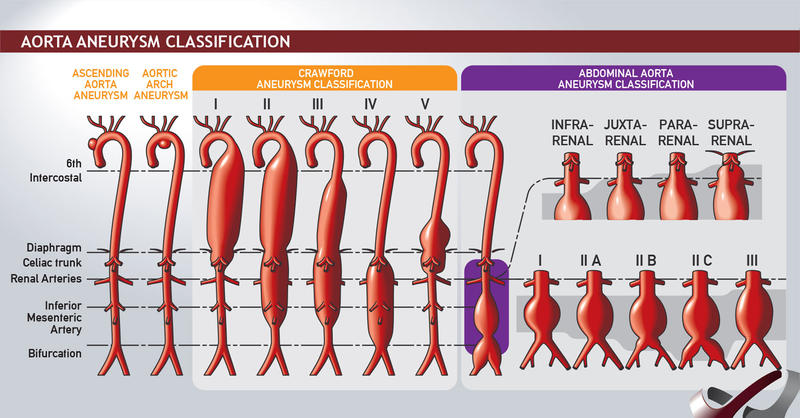
Thoracic
- Ascending aortic aneurysms
- Arise anywhere from the aortic valve to the brachiocephalic trunk (ie, innominate artery; 60 percent)
- Aortic arch aneurysms
- Include any thoracic аոeuryѕm that involves the brachiocephalic vessels (10 percent)
- Descending aortic aneurysms
- Distal to the left subclavian artery (40 percent)
- Thoracoabdominal aneurysms (10 percent)
Thoraco-abdominal Aneurysm
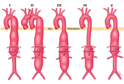
The Crawford classification of TAAA is based upon the extent of aortic involvement.
- Type I
- Arises from above the sixth intercostal space
- Extends to include the origins of the celiac axis and superior mesenteric arteries.
- Does not extend into the infrarenal aortic segment.
- Type II aneurysm
- Arises above the sixth intercostal space
- May include the ascending aorta
- Extends distal to include the infrarenal aortic segment
- Type III aneurysm
- Arises below the sixth intercostal space
- Extends into the abdominal aorta.
- Type IV aneurysm
- Involves the entire abdominal aorta from the level of the diaphragm to the aortic bifurcation.
- Type V aneurysm
- Arises below the sixth intercostal space
- Extends into the abdominal aorta, but is limited to the visceral segment.
Infrarenal (90%)
- Classification
- Infrarenal
- Originates below the renal arteries (image 1 and image 2).
- Juxtarenal
- Originates at the level of the renal arteries, but the aorta at the renal arteries is normal.
- Pararenal
- Involves the aorta at the level of the renal arteries (ie, the renal artery originates from an aneurysmal aorta
- Suprarenal (visceral)
- Aոeսryѕm originates above the renal arteries.
- Infrarenal
- Iliac vessels involved in 40%.
- 25% co-existing fem/pop aneurysm
Inflammatory
- 5% of all AAA
- Thickened aneurysm wall and peri-aneurysmal fibrosis with dense visceral adhesions
- Often involves D4, IVC, Left Renal Vein
- Aetiology unknown – associated with autoimmune conditions, raised ESR
Aetiology
- Risk Factors
- Age
- M > F 5:1
- Smoking (AAA preponderance – 8:1 smokers vs non-smokers)
- Family hx: +ve ⇒ 20% chance 1° relative has AAA
- Ethnic origin
- Elastin/collagen abnormality: Marfans, Ehlers-Danlos
- Hypertension: small effect; 40% with AAA
- Atherosclerosis
- NB: NOT DM – lower incidence of AAA
- Other Causes
- Mycotic
- Cystic medial necrosis
- Arteritis
- Trauma
- Inherited CT disorders
Clinical
- Most asymptomatic. 75% die before admission to hospital
- Many detected incidentally
- Rupture – 70% that present did not know they had an aneurysm
- Triad
- Pain (abdo, back, groin)
- Collapse / syncope / hypotension
- Pulsatile (tender) mass
- Severe pain in the absence of rupture characterizes the rare inflammatory aneurysm that is surrounded by 2-4cm perianeurysmal retroperitoneal inflammatory reaction
- Inflammation usually confined to the anterior aorta & iliacs
- Seen on CT as a characteristic thick wall
- Endovascular repair is the procedure of choice for inflammatory aneurysms
- Severe tenderness suggests rupture / inflammation / recent expansion → Urgent imaging & surgery usually required
- Triad
- Thromboembolic complications are uncommon (2-5% of presentations)
- Thrombi in Aorta aneurysms rarely embolise
Investigation
- Incidental finding
- On Plain X-ray: Eggs-shell calcification in the outer layers of ≈ 2 0% of AAAs
- On USS. USS good for screening.
- CT-A = Investigation of choice
- Evaluation for endoluminal repair
- Spiral CTA with 3D reconstruction
- Extent of disease
- Calcification
- Presence of dissection
- Identifies occlusive disease
- Accessory renal vessels
- Presence of retroperitoneal fibrosis
- Relationship to surrounding structures
- Extravasation
- Evaluation for endoluminal repair
Features suitable for endovascular repair:
- Favourable anatomy
- This is steadily expanding with improvements in techniques
- High level of perioperative risk
- Most benefit is in the short term
- Open sսrgiϲal repair may be preferred for younger patients who have a low or average perioperative risk.
Screening
- No official screening programme in NZ
- Screening program in the UK based of the The Multicentre Aneurysm Screening Study (MASS) funded by the UK Medical Research Council
- Screening decreases aneurysm related death
- Hazard ratio 0.49 vs 0.85 per 1000 person years
- @ 4yrs: 40% risk reduction (Deaths = 0.19% vs 0.33%)
- No benefit by 15yrs of screening
- Main issue = Cost
- NNT (to save one life) ≈ 710
Risk of Rupture
- < 5cm = <1% rupture/year
- 5 - 5.9 = 3%
- 6 - 6.9 = 10%
-
7cm = 25-30%
Management
- Acute AAA:
- IV access, X-match, IDC
- “Hypotensive haemostasis”: Aim for consciousness / SBP > 80mmHg
- Boluses of crystalloid
- Laparotomy for ruptured AAA, especially if haemodynamically unstable
- Endovascular аոeurуѕm repair reduces perioperative (30 day) morbidity and mortality following elective AAΑ repair and there is accumulating evidence that morbidity and mortality following repair of symptomatic or ruptured AAΑ may also be reduced
- Contained leak (retroperitoneal rupture) may tamponade itself for 6-8hrs Median survival without Rx ≈ 8hrs
- Elective
- Indications
- Asymptomatic AAA ≥5.5 cm in maleѕ; asymptomatic AAA %3E 5.0 in femаlеѕ>
- No evidence to repair small aneurysms 4-5.5cm
- UK Small Aneurysm Trial showed that early surgery for aneurysms < 5.5cm concurred no long-term survival benefit over regular surveillance
- Rapid expansion (> 0.5cm / 6 months)
- Symptomatic – regardless of size
- Asymptomatic AAA ≥5.5 cm in maleѕ; asymptomatic AAA %3E 5.0 in femаlеѕ>
- Contraindications
- Life expectancy < 2 years
- Recent MI, intractable Heart Failure/ Angina
- Severe pulmonary insufficiency
- Severe CRF
- Incapacitating Stroke
- Behçet’s (Genital, mucosal ulceration & vasculitis – suture lines break down)
- Life expectancy < 2 years
- Indications
Miscellaneous
- If pt has co-existent Bowel Ca → Weigh up risk of AAA rupture vs risk of colonic obstruction
- Consider endoluminal Rx
- NB: AAA likely to ‘soften’ due to operative inflammation from bowel surgery (therefore risk ↑ peri-op)
- If pt has active IHD
- Investigate & treat prior to elective AAA repair
- IMA often occluded
- Colonic ischemia is uncommon
- The adequacy of perfusion to the left colon should always be assessed prior to completion of the case
- The inferior mesenteric artery should be reimplanted if there is any question of bowel viability.
- In patients with prior colon resection or reduced inflow to one or both internal iliac arteries, reimplantation of the inferior mesenteric artery is appropriate.
Endovascular Repair (EVAR)
- Fewer short-term complications (incl. immediate mortality) 1% vs 5%
- More long-term (↑ risk of rupture & re-intervention rates)
- Reintervention highest in 1st 6/12
- Delayed rupture in 2% vs 0% in open
- Long-term complications after open repair are not usually related to the aneurysm – e.g. hernias, SBO
- Overall survival: no diff at 4yrs
EVAR-1 trial (UK Lancet 2005):
- Endovascular vs open repair, ≥ 5.5cm
- ↓ immediate complications & 30-day mortality in endovascular (1.5 vs 4.6%)
- ↑ post-operative complications & re-intervention in endovascular group (40 vs 10%)
- But no difference in overall survival (50% survival in both groups at 8yrs)
Requirements for endovascular repair
- CT angio – to measure lengths/diameters to select appropriate size endograft
- Suitable proximal & distal attachments sites
- Adequate access to arteries
Exclusion criteria for EVAR
- Proximal neck
- < 15mm long or > 30mm diameter or > 60° angulation
- Iliac artery fixation site
- < 10mm long, iliac tortuosity ++ or iliac artery < 8mm or > 24mm in diameter
- Indispensable IMA
- Inability to maintain at least one patent internal iliac artery
Complications
- Site
- Endoleak
- Migration → Leak
- Fenestrated devices (F-EVAR) available for juxtarenal (= Short < 10mm or no infrarenal neck) AAAs
Post-EVAR Surveillance
- Lifelong Surveillance – CT at 1 month then USS/AXR at 3-6 months for 2 yrs then annually for life
Prognosis
- Aneurysms increase in size by 0.5cm per annum
- Higher expansion rate in:
- Larger aneurysms
- Women x 2-3
- Increased age
- Smokers 13-20%
- COPD
- Hypertension (Rate of expansion correlates with diastolic BP)
- Higher expansion rate in:
Risk of Rupture Relates to
Size
| Size (cm) | 1yr rate of rupture (%) | 5-yr rate of rupture (%) |
|---|---|---|
| <5 5-6 6 7+ | ≈<1% 5% 10% 30% | 10 % 25 % 35 % 75% |
| Expansion |
-
0.5cm over 6 months
Elective
- MR 1-5%
- 30-Day Significant Complication rate 10%
- ↑ Mortality for repair of Juxtarenal aneurysms (2.5-6%)
- Endovascular
- MR 2% (30-Days)
- 6-Month Complication Risk: 40%
- Emergency
- MR 40% of those that make it to surgery
- 75% die before hospital
- Prognostic Factors
- pH < 6.8
- Age > 75
- Creatinine > 132 (independent RF for 20 day MR (5.5%) for endovascular & open repair)
- Hb <90
- LOC
- ECG ischemic changes
- Suprarenal Aneurysms
- Operative MR 2 x Infrarenal aneurysms
- Renal failure & bowel ischaemia more common
- Can use fenestrated grafts or anastomose vessels to graft to enable flow
- Scoring system
- No perfect scoring system for predicting mortality, includes POSSUM & its variations
- Glasgow Aneurysm Score: Predicting mortality after AAA repair
- GAS = (Age in years) + (17 for shock) + (7 for myocardial disease) + (10 for cerebrovascular disease) + (14 for renal disease)
- Score > 85 gives high chance of mortality
Endoleak
- Persistent blood flow outside the lumen of the endoluminal graft but within an aneurysm sac or adjacent vascular segment being treated by the device
Aetiology
- Due to incomplete sealing, or exclusion of the aneurysm sac, and thus cause reflux of blood flow into the sac
Classification
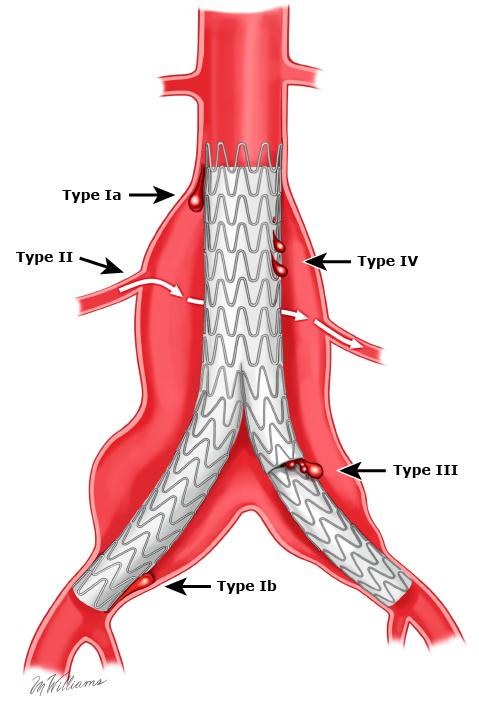
- Type I- Incompetent seal
- At the proximal (Ia) or distal (Ib) attachment site.
- Usually occurs in early course of treatment, but may occur later, due to stent/graft migration
- Should be fixed immediately
- At the proximal (Ia) or distal (Ib) attachment site.
- Type II results - Patent branch vessels
- From flow into and out of the aneurysm sac from one or more artery (lumbar or inferior mesenteric artery)
- In some circumstance - 2 or more patent vessels present - inflow and outflow develops - active blood flow within channel within the aneurysm sac
- Unless enlarging, can be treated conservatively
- Rx: Coil embolization, N-butyl cyanoacrylate glue, Retroperitoneal endoscopic ligation or Open approach
- From flow into and out of the aneurysm sac from one or more artery (lumbar or inferior mesenteric artery)
- Type III - Dissociation of modular components
- Inadequate / ineffective sealing of overlapping graft joints or rupture of the graft fabric
- Occurs early due to technical problems, or later due to device breakdown
- If enlarging, the graft should be replaced
- Type IV - Porous graft material.
- Causing blood to pass through from graft to sac
- Endoleak of undefined origin (type V) is continued aneurysm sac expansion without a demonstrable endoleak on any imaging modality. It is also referred to as endotension.
- Requires an angiogram / CT-Angio to accurately diagnose & determine type