Section: Breast Curriculum: Curriculum, page 9
Definition
- Distension and shortening of subareolar ducts with fibrosis and inflammation
Aetiology
- Ductal Obstruction and Stasis
- Age-related degeneration of ductal epithelium leads to loss of elasticity and ductal dilation.
- Accumulation of secretions causes ductal stasis, which promotes inflammation.
- Periductal Inflammation and Fibrosis
- Stasis triggers chronic inflammation, often with plasma cell infiltration (hence the term plasma cell mastitis).
- Lipid-laden macrophages and lymphocytes infiltrate, further worsening ductal damage.
- Fibrotic Remodeling and Ductal Wall Thickening
- Chronic inflammation leads to fibrosis and scarring around the ducts.
- Fibrosis can cause nipple retraction or a palpable subareolar mass, mimicking malignancy.
- Secondary Infection and Abscess Formation (in some cases)
- Superimposed bacterial infection (commonly Staphylococcus or Streptococcus species) can lead to abscess formation.
- This may present with pain, erythema, and purulent nipple discharge.
- Pts with predominant inflammation (rather than ectasia) tend to be younger
- NOT related to pregnancy or lactation
- Postmenopausal; median age 43, incidence increases with age
- Strong association with smoking
Clinical
- Can present as nipple discharge, retraction, or a palpable mass
- Discharge commonly multifocal &/or bilateral, thick yellow discharge (toothpaste-like)
- Can be clear, white, yellow, grey, green, brown, blue
- Can get associated fistulae
Mammography
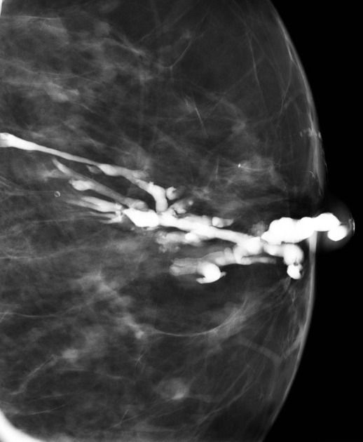
- dilated linear branching densities in subareolar region
- variably present rod-like calcifications pointing towards the nipple
Histology
- Duct dilatation with normal cellular architecture, periductal inflammatory infiltrate (Plasma cells, lymphocytes, PMNs, Giant Cells, Granulomata)
- Bacteria – particularly anaerobes
Management
- As per Nipple discharge
- If confident with the diagnosis = reassure