Section: Breast Curriculum: Curriculum, page 9
Definition
- Benign breast mass (not tumour as does not arise from single cell!)
- Develop from entire lobule
- Both glandular (epithelial) and stromal (connective tissue) components from periductal intralobular fibroblasts
- “Benign fibro-epithelial lesion which originates from both the glandular and stromal components of the breast”
Incidence
- 10% females
- Most common in 21-25 yo - 60% of breast lumps in 20 yr olds
- Incidence decreases after menopause (< 5% occur in women > 50yrs)
- Except HRT
- Account for 1/3 - ½ of breast biopsies for benign disease
- Second most common mass in breast after carcinoma
Classification
- Simple
- Complex
- Calcifications, cysts > 3mm, mixed histological features
- Giant fibroadenoma = > 5cm
- Juvenile – tends to be more cellular (~ puberty)
Aetiology
- Unknown (? ↑ Sensitivity to oestrogen → hyperplasia; ? a continuum of ANDI changes)
- Linked to OCP
- Cyclosporin → Multiple Fibroadenomas
- EBV
- Clinical
- 80% are single
- Hormonally responsive
- Very mobile “breast mouse”
- Origin is from lobular stroma – thus, not tethered to ducts – thus, mobile
- Usually 1-3cm lump
- Non-tender
- Predominate in upper outer quadrant
Pathology
- Part of ANDI complex
- Well circumscribed nodule, pseudoencapsulated
- Proliferation of normal breast elements:
- Intralobular stroma
- Myxoid change, hyaline change
- Stroma often distorts and compresses epithelial components
- Epithelium (± Epithelial hyperplasia ± apocrine metaplasia)
- Intralobular stroma
- “Intracanalicular” & “Pericanalicular” patterns
- Pericanalicular: fibrous tissue surrounding a few small tubular glands; tend to be small & hard
- Intracanalicular: contain more glands, which are indented by fibrous tissue and stretched into elongated, spidery shapes; usually larger & softer
- Low cellularity
- Low mitotic rate
- ‘Complex fibroadenoma’
- ↑ Cellularity, apocrine metaplasia, epithelial hyperplasia
- Differential Diagnosis:
- Cyst, Phyllodes (esp. if > 3cm), Cancer, Hamartoma, Tubular adenoma (entire lesion consisting of glands with very little intervening stroma)
Investigations
- Mass = Triple assessment
- USS
- Well circumscribed
- Wider than it is long
- Not traversing tissue planes
- Homogeneous, (complex fibroadenoma may be heterogenous)
- Mammogram if > 35yrs
- Smooth, density ≥ Surrounding breast (DDx = Cyst, but tend to be perfectly round)
- +/- Stippled calcification with ↑ age
- Popcorn appearance – lobulated appearance
- Classically, fibroadenomas revealed on 1st mammogram
- Core biopsy preferred
- May not need it in a small FA in women < 25 with classical features on imaging
Age of diagnosis: normal breast development stops at around age 25 Thus, if a truly new breast lesion develops after age 25, biopsy is required because it is bizarre for a new fibroadenoma to develop AFTER breast development has completed
Management
- Excise if > 3cm, growing, pt choice, strong FHx, can’t get FNA/core
- 90% of women elect to have them removed
- Surgical treatment = Enucleation
- Same for simple or complex
- NB: One does not need to close the space left behind after removing a fibroadenoma – as the lesion grows, it displaces the adjacent normal breast tissue away. Thus, after removal, the adjacent breast tissue springs back and fills the gap.
- Natural Hx:
- Growth phase, can double in size every 6-12 months
- Show hormonal dependence
- Lactational changes & involution in menopause
- Usually growth ceases at ≈ 2cm in size
- 30% disappear, 50% stay the same, 10% increase, 10% shrink
- Risk of developing Cancer is small (and Lobular more common than Ductal)
- Simple fibroadenoma NO increased risk
- Complex fibroadenoma: RR = 3.5
- Unclear if they can progress to Phyllodes tumour
- Follow-up
- For conservatively treated fibroadenoma, can R/V in 3-6/1
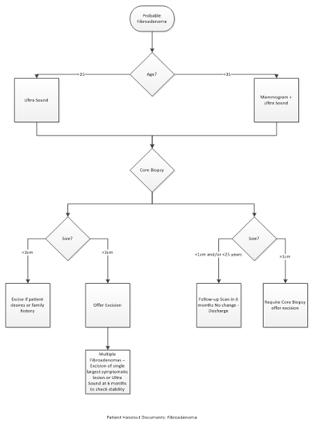
Northshore Hospital flow diagram
- Age
-
35 → USS + Mammogram
- <35 → USS
-
- All get Core biopsy
- Management
- <1cm
- Follow up scan in 6 months, if no change discharge
- <3cm → excise if patient patient desires or family history
-
3cm → offer excision
- <1cm