Section: Breast Curriculum: Curriculum, page 9
Definitions
- Post-lactational involution
- Breast undergoes massive cell death and tissue remodeling as it returns to the pre-pregnant state
- Lobular involution
- Breast epithelial tissue is gradually lost with aging
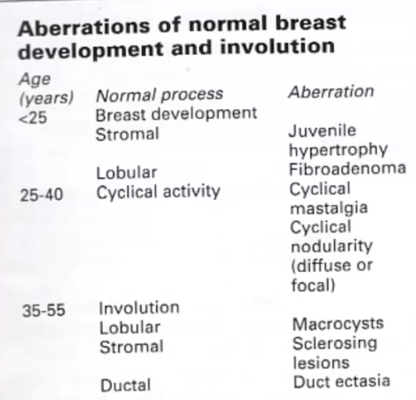
Fibrocystic Change
- Definition
- Fibrosis, Cystosis, Adenosis & Epitheliosis
- Exaggeration of normal hormonal process
- Incidence
- 20-40 years
- 40% of breast lumps are fibrocystic
- 7% women may be symptomatic with cysts
- Aetiology
- ? Minor hormonal imbalances
- Oestrogen > Progesterone, COC protective
- Clinical Manifestations
- Palpable/non-palpable lumps
- Single or multiple (> bilateral)
- Painful – usually sclerosing adenosis with trigger zone
- Recurrent or fast-growing cysts
- Ix:
- Triple assessment if palpable or if other risk factors (e.g. FHx, age, bloody)
- USS & mammogram
- USS shows a solid component → USS-guided Bx
- Atypical features (septations, irregular, solid component) → core / excise
- Cysts aspirated:
- If fluid blood-stained → Triple assessment
- If any residual lumps → Triple assessment
- Cyst fluid typically not sent for cytology as yield is very low and unreliable
- Mx
- If painful mass – Aspirate ± can excise; otherwise mastalgia advice
- Recurrent cysts can be treated with repeat aspiration
- If cyst resolves after aspiration (& screening up to date) reassure and discharge
- Non-palpable cysts are not routinely drained radiologically
- Natural Hx:
- Variation of normal
- 10% of cysts aspirated to dryness recur
- 50% of pts with cysts will develop further cyst over 10 years
- Follow-up 3 months
Fibrocystic Change - Fibrosis
- Overgrowth of fibrous stroma
- Often accompanied with cystic change
- “Fibrocystic change”
- ↑ fibrous stroma with duct dilatation & formation of cysts
- ? may arise as a result of cyst rupture → Chronic inflammation → Fibrosis
Fibrocystic Change - Cystosis
- Cyst = Fluid filled cavity lined by epithelial cells
- Cystic involution of the breast lobule (Apocrine)
- Outflow obstruction due to epithelial hyperplasia / stricture or fibrosis of terminal ductules / kinking of ducts + secretion by distal lobule → Cyst
- Lobules develop microcysts → coalesce into larger cysts
- Characteristic halos on mammography
- USS essential
- Can distinguish solid and cystic lesions
- Also give info on septations and differentiate simple and complex cysts
- USS essential
- Simple Breast Cysts
- No association with cancer
- Lined by simple cuboidal epithelium
- Classic USS features meaning likely benign
- Wall calcified
- Mobile particles (usually cholesterol crystals)
- Complex Breast Cysts
- Possible association with cancer
- Lined by apocrine epithelium (apocrine metaplasia) with columnar cells
- High K:Na ratio
- Tend to recur
Cancer risk ↑ed (estimated >2%) if:
- Blood in cyst
- Lump present after aspiration
- Thickened walls
- Thick (> 2mm) internal septations
- Mix of cystic & solid components
- Co-existent Florid ductal hyperplasia
Fibrocystic Change - Adenosis
- Increase in number of lobular acini / glandular elements
- Cells & basement membrane have normal relationship
- 3 types:
- Sclerosing adenosis
- Proliferation of terminal duct lobules (enlargement & distortion)
- ↑ no. of acini + fibrous stromal ∆ with intralobular fibrosis
- ± Associated with microcalcifications
- Differentiated from Radial Scars (RS)/ Complex Sclerosing Lesion (CSL) by degree of excessive myoepithelial proliferation seen in RS and CSL
- Mx - Nil
- Radial Scar (< 1cm)
- Complex Sclerosing Lesion (> 1cm)
- Background for development of atypical epithelial proliferations including ADH (Atypical Ductal Hyperplasia) and DCIS
- Increased risk of malignancy
- Difficult to distinguish from malignancy – associated with malignancy ~ 10%
- Mx - Open excision recommended
- Sclerosing adenosis
Fibrocystic Change – Epithelial Hyperplasia
- Usually incidental finding
- Proliferation of epithelial cells in ducts or lobules with ≥ 3 layers of cells
- Normal = 2 cell layers (epithelial & myoepithelial)
- Hyperplasia: ↑ed no. of layers ± ‘fenestrations’ within layers
- Classification: Ductal vs. Lobular ± Atypia
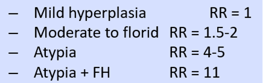
- Path: extent of hyperplasia:

RR of Cancer: