Section: Breast Curriculum: Curriculum, page 10
Definition
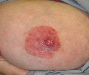
Paget’s disease of the nipple is a breast cancer that begins in the nipple and can extend to the surrounding areola. It is characterized by malignant cells (Paget cells) infiltrating the epidermis of the nipple, often associated with an underlying ductal carcinoma, either in situ (DCIS) or invasive breast cancer.
- Eczematous inflammatory condition, particularly involving nipple & areola
- Typically the epidermal manifestation of an underlying cancer
- 95% have underlying cancer
- Two characteristic findings
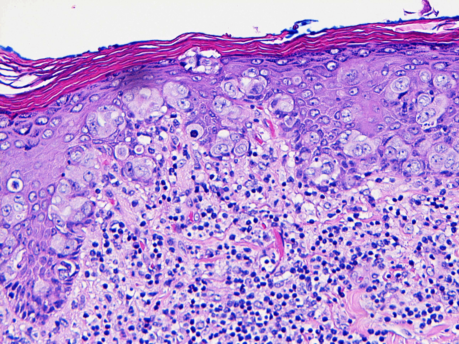
- Pagetoid Cells
- Large, round cells with pleomorphic nuclei + abundant clear cytoplasm
- Reactive changes in epidermis
- Lymphocyte infiltration + Angiogenesis
- Pagetoid Cells
Incidence
- Uncommon; associated with ≈ 1% of all Breast Cancer
- Occurs most commonly in post-menopausal
- More common in Polynesians
Classification
- Paget’s disease NOS
- Paget’s with DCIS
- Paget’s with invasive ductal cancer
Clinical
- Eczema / dermatitis changes of nipple-areolar complex
- Begins in nipple and migrates out towards areolar
- NB: Opposite to eczema which starts in areola
- Crusted, oozy patches of skin, but distinct edges, not itchy no vesicles
- Ulceration, discharge, bleeding, fissures & surrounding inflammation
- Unilateral
- Rarely involves the skin of the breast
- Associated with a palpable mass in up to ≈ 50%
- Mass represents cancer
Aetiology / Pathology
-
Epidermotropic Theory (more widely accepted)
- Cells migrate along the BM and enter the epidermis and dermis of the nipple areolar complex
- Pagetoid cells and underlying malignant cells demonstrate the same IHC pattern
-
Transformation Theory (fallen out of favour)
- Paget Disease arises from epidermal keratinocytes (Toker Cells) independent of underlying breast malignancy; actually represents epidermal carcinoma in situ
- Hypothesis: Breast duct secretions continuously damage epithelium leading to malignant transformation of keratinocytes
-
Underlying DCIS or invasive ductal Cancer Typically poorly differentiated 90% of Paget cells are HER-2 positive
Microscopic
- Two characteristic findings
- Pagetoid cells
- The nipple epidermis becomes infiltrated with “paget cells”
- Large, pale staining, abundant cytoplasm and round/oval nuclei
- Scattered between the keratinocytes
- Like in DCIS, the cells do not infiltrate beyond the BM
- The nipple epidermis becomes infiltrated with “paget cells”
- Reactive changes in epidermis and dermis with lymphocytic infiltration and angiogenesis
- Pagetoid cells
- IHC stain for CK7, CAM-5.2, AE1/AE3 and S100.
- 90% of Paget cells are HER2 +ve
Investigations
- Confirm Paget’s disease
- Full-thickness nipple-areolar complex biopsy or excision biopsy if non-diagnostic
- Then look for underlying malignancy
- Triple assess
- Mamm less sensitive in setting of Paget’s with only 30% detection rate, therefore if no malignancy on mamm consider MRI
Management
- Surgical
- Resection of nipple/areolar complex a minimum
- Treatment of underlying Breast cancer & resection of nipple-areola complex
- Mastectomy
- Usually mastectomy (high frequency of multicentric disease)
- BSC
- Can consider WLE + Radiotherapy if central disease only
- When no associated lesion is found – treat as you would DCIS
- Resect nipple-areola complex with at least a 2cm cone of retroareolar tissue: WLE + RTx or mastectomy
- ? SLNBx
- Incidence of axillary mets ≈ 12%
- Do if confirmed cancer or planning Mastectomy
- Adjuvant treatment
- As per the underlying cancer
- No evidence for endocrine therapy in women with PDB without an associated invasive cancer or DCIS
Prognosis / Natural Hx:
- Tumour stage is the best predictor rather than the presence of Paget disease per se
- When lesion consists of nipple changes only, the incidence of axillary mets is <5%, when a breast mass is also present, the incidence of axillary mets ↑s
- After cone excisions of nipple-areolar complex → ≈ 40% recurrence at up to 5yrs
Follow-up
- Breast Ca surveillance