Section: Colorectal Sub-section: Colorectal cancer Curriculum: Curriculum, page 21
Natural History
- 50% left sided, 25% right sided, 30% rectum
- Epidemiology: 2nd most common cancer in NZ.
- Average age of adenomas ~5 years younger than carcinomas.
- Large adenomas more likely to display cellular atypia and genetic abnormalities than small lesions
- 20% of patients with colonic cancer will present as an emergency and 16% will present with obstruction.
- Ways of spread:
- Direct spread
- Lymphatic
- Haematogenous
- Transcoelomic – peritoneum
Anatomy
There are four layer: Mucosa
- Epithelium
- Lamina propria
- Muscularis mucosa Submucosa Muscularis propria
- Inner circular
- Outer longitudinal Aventia or serosa
Embryology
Aetiology
Non modifiable:
- Genetic factors
- Lynch syndrome
- FAP
- Any family history Predisposing conditions
- Ulcerative colitis
- Crohn’s
- Previous gastric surgery – possibly due to altered bile acid metabolism
Modifiable:
- Diet – low fibre, low intake of vegetables, red meat, alcohol, fat and sugar
- Lifestyle – decreased physical activity, obesity, smoking
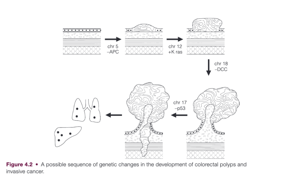
Genetic factors in sporadic colorectal cancer
- APC gene mutation - adenomatous polyposis coli gene
- central to ordered cell motility, are thought to occur early as they are found in 60% of all adenomas and carcinomas.
- K-ras mutations
- induce cell growth by activating growth factor signal transduction.
- p53 gene mutation
- common in invasive colonic cancers but rare in adenomas and is therefore deemed to be a late event that accompanies the development of invasion.1
- p53 protein has roles in the repair of DNA and the induction of programmed cell death
Clinical presentation
Emergency
- Obstruction
- Bleeding
Chronic symptoms
- Anaemia
- Change in bowels
- Abdominal pain
- PR bleeding – mostly rectal
- Tenesmus – mostly large rectal
- Mass
- Pneumaturia
- Vomiting
Screening
- In NZ 60 – 74. Māori and Pacific from age 50 in some parts of NZ.
- WHO principles of population screening
- Important health problem
- Suitable latent asymptomatic stage
- Natural history of condition understood
- Acceptable treatment
- Facilities for diagnosis and treatment
- Suitable test - high specificity and sensitivity
- Safe and acceptable to population screened
- Agreed policy on who to treat
- Cost of finding is economically balanced with overall health
- Case finding should be continuous process
- Biases in screening:
- Selection bias – chooses people more health conscious
- Length bias – tendency for screening to detect disproportionate number of cancers that are slow growing with good prognosis.
- Lead-time bias – time from date of detection by screening and time it would have been detected if not screened. Given survival measured from date of diagnosis, screening advances the date without necessarily prolonging survival.
- Modalities
- iFOBT (Immunochemical Faecal Occult Blood Test)
- gFOBT (Guaiac Faecal Occult Blood Test)
Screening groups
Colonoscopy guidelines in NZ for asymptomatic patients with a FHX.
- Group 1 – low risk
- Group 2 – moderate risk
- Group 3 – high risk.
Group 1
- Patients with first degree relative who had CRC > 55 years old.
- There is no specific surveillance recommendations for this group.
Group 2
- One first degree relative diagnosed with CRC < 55 years.
- Two first degree relatives on the same side of the family diagnosed with CRC at any age.
- Should have colonoscopy 5 yearly from aged 50 OR 10 years prior to earliest age at which CRC was diagnosed in family member. Once aged 60 – can join NBSB
Group 3
- Fhx of Lynch, FAP, or other CRC syndromes.
- 3 relatives on same side of family having CRC at any age with one being a first degree relative.
- There are quite a few other criteria but generally this looks Lynch syndrome risk factors and polyposis syndrome risk factors.
- These patients should be referred to the NZFGCS
Investigations
- Colonoscopy/sigmoidoscopy
- CT – CAP for staging
- CT colonography – polyps down to 6 mm
- MRI rectum
- Good correlation between extramural spread of tumour seen on MRI and histopathology. Can identify when tumour extends to or within 1 mm of mesorectal fascia (CRM) (MERCURY study).
- Can also predict vascular invasion and LN involvement but sensitivity and specificity for nodal status is suboptimal.
- Endorectal US – used in some centres for T staging of early rectal cancers
- PET-CT – limited role, recommended when surgical resection of metastases considered to exclude occult disease.
Histology
Macroscopic description
- Size of tumour
- Site of tumour in relation to resection margins
- Any abnormalities of background bowel
Microscopic description
- Histological type
- Differentiation based on predominant grade
- Completeness of excision at cut ends, including doughnuts and at any radial margin
- Presence of extramural vascular invasion
- Pathological staging according to Dukes’ or TNM
- Maximum extent of invasion through bowel wall
- Serosal involvement by tumour
- Number of lymph nodes and whether apical node involved.
Most common subtypes of colorectal adenocarcinoma
- Mucinous adenocarcinoma – 10% of all adenocarcinomas. >50% of the tumour volume is composed of extra-cellular mucin. Poorer prognosis
- Signet ring cell adenocarcinoma - < 1%. Requires >50% of tumour cells to show signet cell features. Often poorly differentiated high grade tumours. Associated with Lynch
- Medullary carcinoma – sheets of epithelioid neoplastic cells with a pushing border and tumour infiltrating lymphocytes. Associated with Lynch. Better prognosis.
Grading
- Is based on percentage of tumour which is glandular.
-
95% glandular – well differentiated – Grade 1
- 50-95% - moderately differentiated – Grade 2
- < 50% - poorly differentiated – Grade 3
Staging
| Dukes’ staging | |
|---|---|
| A | Invasive carcinoma not breaching muscularis propria |
| B | Invasive carcinoma not involving regional nodes |
| C1 | Invasive carcinoma involving regional nodes, but not apical node |
| C2 | Invasive carcinoma involving regional nodes, but apical node positive |
| D | Widespread metastases |
| TNM staging - AJCC 8th edition |
| TNM staging | ||
|---|---|---|
| T1 | Tumour invades submucosa. Tis is it has not grown beyond the mucularis mucosa | |
| T2 | Tumour invades muscularis propria | |
| T3 | Tumour invades subserosa or into non-peritonealised pericolic or perirectal tissues | |
| T4 | Tumour perforates visceral peritoneum (a) or directly invades other organs or structures (b) | |
| N1 | Mets in 1-3 pericolic or perirectal lymph nodes. 1 (a), 2-3(b) | |
| N2 | Mets in 4 or more pericolic or perirectal nodes. 4-6 (a), >6(b) | |
| M1 | Distant mets. One organ (a), more than one organ (b), peritoneum (c) |
Rectal cancer T0: no evidence of primary tumour Tis: carcinoma in situ: intraepithelial or invasion of lamina propria T1: tumour invades submucosa T2: tumour invades muscularis propria
- MRI does not yet have the resolution capable of enabling differentiation of T1 and T2 lesions T3: tumour invades through the muscularis propria into the subserosa or into non-peritonealised perirectal tissues without reaching the mesorectal fascia or adjacent organs
- T3a: tumour extends <1 mm beyond muscularis propria
- T3b: tumour extends 1-5 mm beyond muscularis propria
- T3c: tumour extends 5-15 mm beyond muscularis propria
- T3d: tumour extends 15 mm beyond muscularis propria T4: tumour invades directly into other organs or structures and/or perforates visceral peritoneum
- T4a: tumour penetrates to the surface of the visceral peritoneum
- T4b: tumour directly invades or is adherent to other organs or structures
Nx: regional nodes cannot be assessed N0: no regional lymph node metastases N1: metastasis in 1-3 regional (perirectal) lymph nodes
- N1a: metastasis in 1 regional lymph node
- N1b: metastasis in 2-3 regional lymph nodes
- N1c: tumour deposit(s) in the subserosa, mesentery, or non-peritonealised pericolic or perirectal tissues without regional nodal metastasis N2: metastasis in 4 or more regional lymph nodes
- N2a: metastasis in 4-6 regional lymph nodes
- N2b: metastasis in 7 or more regional lymph nodes
Mx: cannot be assessed M0: no distant metastasis M1: distant metastasis
- M1a: metastasis confined to one organ or site (for example, liver, lung, ovary, non-regional node) without peritoneal metastases
- M1b: metastases in more than one organ
- M1c: metastasis to the peritoneum with or without other organ involvement

Stage groupings stage 0: Tis N0 M0 stage I: T1-2, N0 M0 stage II: T3-T4b, N0 M0 stage III: AnyT, N1-2, M0 Stage IV, AnyT, AnyN, M1
Perioperative considerations
- ERAS protocol - meet the nurse prior
- Pre-habilitation with exercise protocol
- Assessment + optimisation of comorbidities: cardiorespiratory testing, sleep testing, respiratory testing, optimise diabetes etc
- Nutritional state - may require a period of feeding or obesity/weight loss
- Smoking cessation
- Stoma marking
- Anaemia – iron or RBC transfusion.
- Preop bowel prep and oral antibiotics
- Picolax®. Evidence is mixed.
- VTE: LMWH for 28 days postop
- Skin cover antibiotics - cephalosporin or ertapenum
ERAS
- Pre-operative eduction
- Appropriate multi-modal analgesia
- Early mobilisation
- Early TROC
- Early drain remove
- No restriction on oral intake and avoidance of NG tube
Complication:
- Anastomotic leak
- Definition: the leak of luminal contents from a surgical join between two hollow viscera diagnosed:
- Radiologically by CT
- Clinically with evidence of bowel contents or gas through drain or wound
- Endoscopy
- Intraoperatively
- Definition: the leak of luminal contents from a surgical join between two hollow viscera diagnosed:
- Severity classification:
- Grade A: no change in patient management
- Grade B: requiring intervention but no relaparotomy
- Grade C: requiring relaparotomy