Sub-section: Adrenal Section: Endocrine
Hyperaldosteronism
Definition
Primary hyperaldosteronism
- Conn’s Syndrome
- Autonomous release of excess aldosterone from one or both adrenal glands
- From Zona Glomerulosa
- Commonest cause of 2° HTN
- 5-13% of all HTN
- Mean age presentation = 50yrs
- M > F (slight)
Secondary Hyperaldosteronism
- Inactivation of RAS mechanism in Liver Failure, HF, Nephrotic Syndrome etc.
Presentation and Sequelae
- Classically manifests as
- Hypertension (unusually only sign)
- +/- Hypokalaemia
- Most have normal K+
- More likely in severe cases/late stage
- Patients often asymptomatic
- May have muscle cramps, weakness, paraesthesias from low K+
- Often moderate/severe hypertension, refractory to medical rx
- Often on 2-4 antihypertensives
- May get responsiveness to spironolactone
- Predictive of good response to surgical treatment also
- Associated with increased risk of:
- Stroke, MI, AF and VF, CHF
- More pronounced than effect of HTN alone
- Surgical removal reverses the risk
Aetiology
- Most commonly
- Bilateral Hyperplasia
- Unilateral Aldosteronoma
- Others (All rare, < 1%)
- Aldosterone-Secreting Adrenocortical Carcinoma
- Unilateral Adrenal Hyperplasia
- Familial Hyperaldosteronism
- Type 1 – Glucocorticoid suppressible
- Type 2 – Unable to be suppressed by glucocorticoids
Diagnosis
- Consider biochemical screening if
- Hypertension and hypokalaemia
- Hypertension alone that is treatment resistant or if < 40yrs old
- Incidentalomas
- Workup
- Screening
- Renin/aldosterone ratio
- ⇧ Plasma Aldosterone concentration (PAC)
- Decreased Plasma Renin activity or concentration (PRA/PRC)
- Net effect is that there is a high PAC:PRA ratio > 20-30
- NB: Aldosterone antagonists, beta-blockers, CCBs, ACEI’s can interfere with results and should be stopped prior
- Confirmation
- Oral sodium loading
- Saline infusion test
- Or less commonly fludrocortisone suppression test
- If no suppression, diagnosis confirmed
- Screening
Localisation
- Preferred initial imaging is fine slice adrenal CT
- Most aldosteronomas are < 2cm
- Tend to be homogenous
- C.f. carcinomas which are heterogenous, often > 4cm
- NB: Can have false +ve
- Primary hyperaldosteronism attributed to a non-functioning adenoma seen on imaging
- Non-functioning adenomas more common > 40yrs
- Primary hyperaldosteronism attributed to a non-functioning adenoma seen on imaging
- Most aldosteronomas are < 2cm
- Adrenal vein sampling
- Standard test to distinguish between unilateral adenoma and bilateral hyperplasia or if bilateral lesions to localise a side for treatment
Adrenal Vein Sampling
- Simultaneously measures cortisol and aldosterone ratios in peripheral circulation and L+ R Adrenal Veins
- Indications:
- If CT normal or has bilateral abnormalities
- If unilateral abnormality and age > 35
- As a young patient is unlikely to have an incidental adenoma
- Can give bolus or infusion of cosyntropin (ACTH)
- Confirm successful catheterization
- Adrenal vein to IVC cortisol ratio 3:1
- Measure Aldosterone/Cortisol ratio
- Accounts dilutional effect of the inferior phrenic vein flow into the left adrenal vein; these are termed “cortisol-corrected ratios.”
- If one side ratio is > 4x the opposite, confirms localisation
- In skilled hands – Sensitivity 95%, Specificity 100%
- NB: Often fails due to inability to cannulate Right Adrenal Vein (success rates 40-80%)
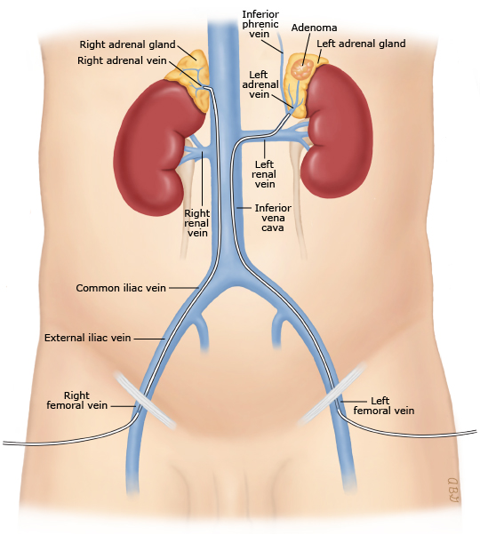
- NB: Often fails due to inability to cannulate Right Adrenal Vein (success rates 40-80%)
Surgical Management
- Correct HTN and hypokalaemia prior to surgery
- If unilateral disease localised
- Minimally invasive Adrenalectomy
- If not surgical candidate
- Aldosterone antagonist – Spironolactone
- Side effects include gynaecomastia, libido loss, menstrual irregularity, erectile dysfunction
- Aldosterone antagonist – Spironolactone
- Need to consider risk of malignancy prior to advocating surgery
- If Bilateral Adrenal Hyperplasia
- Spironolactone
- If Type 1 Familial Hyperaldosteronism
- Steroids
Outcomes after Adrenalectomy for Aldosteronoma
- Cure defined as
- Reduction of hypertension
- Reduction of aldosterone levels
- Resolution of hypokalaemia (if present)
- Cure rates 75-95%
-
80% have normalization of BP or significant reduction in anti- HTN needed
-
- NB: Can have overlap with essential HTN
- BP may not completely normalise
Cushing’s Syndrome
Definition and Incidence
- Cushing’s Syndrome
- Disorder characterised by a no. of signs and symptoms, caused by long term effects of elevated glucocorticoid levels
- Cushing’s Disease
- Glucocorticoid excess caused by an ACTH-secreting Pituitary Adenoma
Clinical Presentation
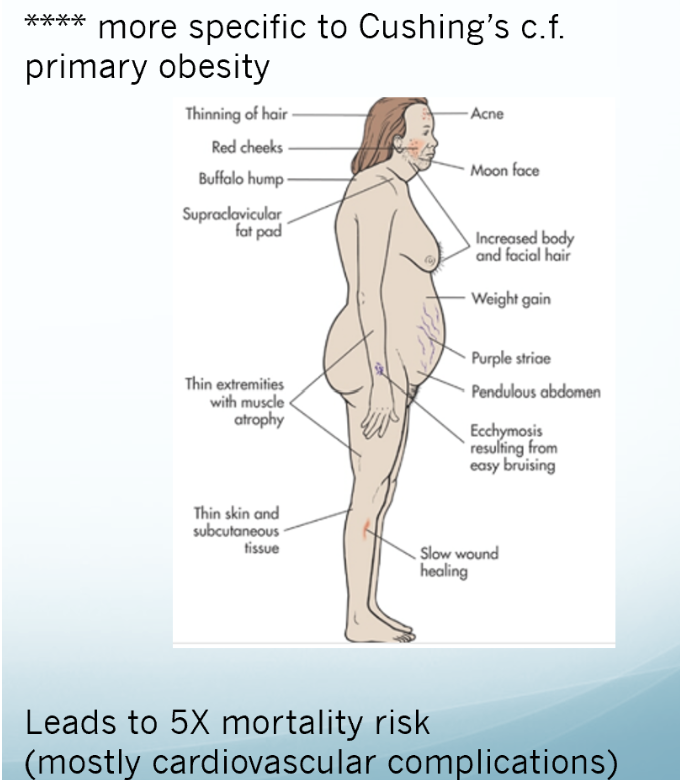
- Symptoms
- Central obesity
- Buffalo hump
- Moon faces
- Facial Plethora ****
- Reddening due to thin skin
- Striae****
- Hirsutism
- Loss of libido
- Menstrual irregularity
- Depression/psychosis
- Proximal myopathy****
- Easy bruising****
- Signs
- Diabetes Mellitus
- Osteoporosis
- Hypertension (> 70%)
- Hypokalaemia
Aetiology
- Most common cause is exogenous steroid use
- Endogenous Cushing’s syndrome is rare (F > M)
- ACTH-Dependent (80%)
- Pituitary-related (Cushing’s disease) (70%)
- Ectopic (10%)
- E.g. NET’s, Bronchogenic Carcinoid, Medullary Thyroid, Phaeo, or Small Cell Lung Ca
- ACTH-Independent (15-20%)
- Primary adrenal Cushing syndrome
- Adenoma (90%)
- Carcinoma (10%)
- Bilateral micronodular/ macronodular Hyperplasia (<1%)
- Primary adrenal Cushing syndrome
- ACTH-Dependent (80%)
Diagnosis
- Screening tests
- 24hr Urinary Free Cortisol
- Reflects average cortisol levels
- Levels > 4x normal - diagnostic of Cushing’s
- Less sensitive at diagnosing subclinical Cushing’s
- Do at least 2 measurements, may need 3
- Late-night Salivary Cortisol
- Cushing’s syndrome shows loss of normal circadian rhythm of cortisol secretion
- Resting midnight plasma cortisol > 50nmol/L is 100% sensitive for Cushing’s
- Salivary results closely correlated to plasma
- High sensitivity and specificity for Cushing’s (> 92%)
- Need to do in low stress environment
- Hence: At home
- Low Dose Dexamethasone Suppression Test
- Exploits loss of negative feedback loop for cortisol
- 1-2mg dexamethasone, given at midnight
- Should suppress cortisol level taken at 9am following am
- If cortisol > 50nmol/L the following am
- Sensitivity for Cushing’s 95%
- Specificity 80%
- Some units may do 2mg/day, 48hr testing
- Gives higher specificity
- 24hr Urinary Free Cortisol
- Establishing a cause
- Corticotropin dependent or ACTH-independent disease
- High ACTH = ACTH-dependent
-
20 pg/mL, 4.4 pmol/L
-
- Low ACTH = ACTH-independent
- <5 pg/mL (1.1 pmol/L
- Intermediate
- Repeat testing
- High ACTH = ACTH-dependent
- ACTH-dependent
- Pituitary vs ectopic
- Noninvasive biochemical testing with
- CRH (if available) or desmopressin (DDAVP) stimulation test
- Both stimulate ACTH
- Increases in ACTH and cortisol is consistent with pituitary adenoma
- CRH (if available) or desmopressin (DDAVP) stimulation test
- High dose dexamethasone suppression
- Pituitary tumours retain some negative feedback control and ectopic source does not
- Give single dose 8mg dexamethasone, or 2mg QID for 48hrs
- If cortisol level drops to < 50% baseline
- Suggests Pituitary Cushing’s
- If no significant drop - Ectopic cause (or Adrenal cause, but will have already ruled this out with prior investigations)
- Imaging
- MRI
- Invasive testing
- Bilateral Inferior Petrosal Sinus Sampling
- To differentiate pituitary Cushing’s from ectopic if dexamethasone suppression test equivocal
- Invasive
- Catheter measures ACTH in each Petrosal Sinus and peripheral ACTH levels
- Central-to-peripheral ACTH gradient
- Catheter measures ACTH in each Petrosal Sinus and peripheral ACTH levels
- Bilateral Inferior Petrosal Sinus Sampling
- Further Investigations of Ectopic ACTH Dependent Cushing’s
- Basic tumour markers
- Urinary 5-HIAA
- Serum Calcitonin
- Chromogranin A
- Other GI neuroendocrine hormones as indicated
- CT neck/chest/abdo/pelvis
- Somatostatin receptor scintigraphy
- Basic tumour markers
- Noninvasive biochemical testing with
- Pituitary vs ectopic
- ACTH Independent
- Adrenal Cushing’s
- Next step is Localisation
- CT is the initial modality of choice
- Will see adenoma, carcinoma, and macronodular hyperplasia
- Won’t see micronodular hyperplasia
- NB: Overlap in appearances of adrenal adenoma and carcinoma on cross sectional imaging
- Risk of malignancy increased if
- High attenuation
- Irregular outline
- Delayed contrast washout
- Heterogenous
-
4cm in size
- If concerned about malignancy:
- MRI with Gadolinium can help to differentiate
- High water content of carcinoma c.f. adenoma
- CT is the initial modality of choice
- Corticotropin dependent or ACTH-independent disease
Management
- Peri- and Post-op glucocorticoid administration
- Essential
- Initial high dose with taper post-op
- Some units withhold initially post-op
- To assess cortisol levels, ? biochemical cure
- Beware of Adrenal Crisis – Endocrinologist should manage
- Some units withhold initially post-op
- Surgery
- Primary adrenal Cushing’s syndrome
- Cushing’s disease
- Ectopic ACTH independent Cushing’s syndrome
Surgical Management of Primary Adrenal Cushing’s
- Unilateral Adrenalectomy
-
90% effective, but resolution of symptoms takes months-to-years
- Failure could be due to missed or recurrent malignant disease
-
- Perform in specialised center
- High risk of infections, Skin injury, Fractures, Hyperglycaemia
- DVT risk increased (up to 5%) so use clexane
Surgical Management of Cushing’s Disease (Pituitary)
- Initial approach
- Pituitary microsurgery for Cushing’s disease
- Trans-nasal, trans-sphenoidal approach
- If fails, consider:
- Re-operation
- Danger of causing pan-hypopituitarism
- Radiation
- If fail
- Bilateral Adrenalectomy
- Will need lifelong glucocorticoids and mineralocorticoid supplementation
Management Ectopic Cushing’s Syndrome
- Treatment of primary tumour is preferable
- But not always possible
- Controlling symptoms paramount if removing primary not possible
- Ketoconazole inhibits cortisol production
- Bilateral Adrenalectomy best way to achieve symptom palliation
- Even in patients with limited life expectancy
- Esp. if laparoscopic
Sex Steroid Excess
- Very rare
- Most virilising (as opposed to feminising)
- 1/3 virilising are malignant
- Almost all feminising lesions are malignant
- 20% of adrenocortical carcinomas cause virilisation
- 24% of adrenocortical carcinomas show mixed virilization and Cushing’s Syndrome
- Tests
- 24hr urine Testosterone, DHEA and DHEAs
- Treatment
- Laparoscopic Adrenalectomy
- High probability of malignancy so close radiographic and intra-op inspection needed
- If any concerns – Open Adrenalectomy