Section: UGI Sub-section: Bariatrics Curriculum: Curriculum, page 83
Slipped gastric band
- Present with obstructive symptoms
- 15% 1st 3 yrs, > 50% at 10 yrs
- Typically at 2-3 yrs
- Usually anterior slippage
- Posterior slippage rare
- Investigations
- AXR to look at PHI angle
- Posterior slippage
- O-sign - gastric band being visible end on
- Anterior slippage – band rotates clockwise
- Recognised by loss of normal phi angle (10 - 60 degrees)
- Posterior slippage
- Contrast swallow
- AXR to look at PHI angle
- Creates acute angle between oesophagus and stomach
- Can lead to ischaemia of Fundus
- Management
- Deflate band completely, may resolve symptoms
- If works, then adequate acute rx, and send back to surgeon for further review
- Deflate band completely, may resolve symptoms
- May need removal of band
- Or repositioning (by bariatric surgeon)


Eroded gastric band
- 1-7%
- Aetiology
- Gastric wall ischaemia from tight band placement
- Trauma/injury during band placement
- Presents with
- Pain, nausea, vomiting +/- haematemesis
- Failure of weight loss/Loss of restriction
- Red port sign (infected port)
- Usually seals itself so unusual to see free peritonitis
- Investigations
- Diagnosed endoscopically
- CT scan to look for leak/collection (rare)
- Management
- Removal of band - Generally lap
- Cut down over the capsule surrounding the band
- There may be gastro-gastric stitches that need to be removed
- Cut down onto buckle of band with diathermy
- Divide band and then remove it
- May need distal gastrotomy to access if phlegmon
- Leave a drain
- ALWAYS – Keep all pieces of the band on a mayo table and reassemble to be sure entire band is removed
- Removal of band - Generally lap


Gastric band obstruction
- Dysphagia
- Can be
- Slippage
- Over inflation
- Fibrotic foreign body reaction/oedema from surgery
- First step is to remove all of fluid from band
- Pillow behind lower back
- Lift head like sit-up
- Brace with fingers and insert Huber needle
- Huber needle is non-coring
- If can’t find, then use 25g fine bore
- NB: Can get pseudo-achalasia with the bands and resultant dysmotility
Post-op Leak
Bypsss
- JJ, GJ, Remant pouch, blind limb
Contained vs uncontained Stable vs unstable
Princples Source control Exclude distal obstruction Abx Nutrition Pyschosoical Treatment injury Communication with family and patient
Porto-mesenteric vein thrombosis
- 1% sleeve
- up to 1m after
Sleeve
- One staple line
- Definition
- Deep/organ space surgical site infection
- Infrequent but can be severe
- Deep/organ space surgical site infection
- Is it
- Localised vs Lateralised?
- Early vs Persistent?
- Stenosis involved?
- Key points
- Stents (Megastent) very handy way of managing
- But don’t over-rely or keep them in too long
- Early and repeated use of endoscopy invaluable
- Assessment, drainage, dilatation, stenting, feeding
- Prevent external fistula by conservative use of external drainage
- And removing any external drains early
- Focus on
- Drainage of cavity, then ablation of the cavity
- Treatment of stenosis
- Rapid discharge
- Conversion of sleeve to RYGB is valid option for leak
- Stents (Megastent) very handy way of managing
Aetiology
- Crossed staple lines
- Anastomotic tension/ Ischaemia
- Stenosis
- Sleeve leaks more common than RYGB
- High pressure organ with sphincters at both ends
- At angle of HIS as this is the thinnest and most avascular part
- High pressure organ with sphincters at both ends
- Most in first week, often after discharge
- Can be weeks, months, years later
- Presentation - Sepsis, pain
- Investigations
- CRP (if high be concerned)
- Contrast swallow CT – 80-90% sensitivity
- Free gas, fluid, +/- contrast extravasation
- Abscess at top of sleeve = Leak until proven otherwise
- Left pleural effusion may be sign of evolving leak
- Fluoroscopy = 70% sensitivity
Classification
- Type 1
- Phlegmon only
- Oedema around staple line, maybe small amount of gas
- Type 2
- Abscess (with or without contrast extravasation)
- 2a Close to staple line
- 2b Extends away from staple line
- Type 3
- Generalised Peritonitis
- Gas and fluid throughout abdomen
- Type 4
- Chronic Fistula
- 4a Close to staple line
- 4b Distant
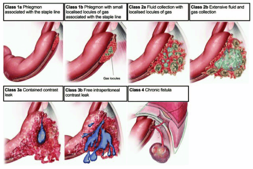
Management of bariatric
Early Leak (Type 1)
- Size of cavity, stenosis, and ileus are the main drivers
- Appropriate resuscitation, antibiotics and nutrition
- Aim for rapid discharge (within 1-2 weeks)
- Avoid surgery if possible
- Avoid resection if surgery done
- If patient does not progress, they are getting worse!
- Try something different
- Phlegmon
- IV Abx, observation
- Fasting until subjectively improved or CRP falling
- Then high protein fluids
- Low threshold for OGD within first 24hrs
Type 2a - Medial Abscess
- Endoscopy and internal drainage (better if available)
- Suitable for sleeve, total gastrectomy, staple-line leak in RYGB
- Avoids fistula formation
- Allows endoscopic reassessment of defect
- Can place NJ feeding tube past it
- Aim for PO fluids day 1, and early discharge
- External Drainage if not available
- Laparoscopy and drainage, or CT guided drainage
- But will create external fistula unless drain removed early
Type 2b - Lateral Acute Abscess
- Usually more complex and unwell
- May have ileus +/- stenosis driving increased intraluminal pressure and increased leak volume
- Patients need rapid sepsis control and drainage
- Laparoscopy, external drainage, and stent (esp. RYGB)
- NB: Laparoscopy will convert small abscess to large abscess
- Stents very good at preventing fistulas but not as good at treating them
- So place stent early!
- Consider endosponge
- Laparoscopy, external drainage, and stent (esp. RYGB)
- Remove stents or endosponges after 2/52 and convert to internal drains
- Usually need TPN
Type 3 - Generalised Peritonitis
- Laparoscopy, lavage, and drain
- On table endoscopy and stent
- Place stent from 4cm above GOJ
- If sleeve, go to pre-or post-pyloric distally
- If RYGB, go across anastomosis
- Lap suture stent to stomach/small bowel to prevent migration
- Place stent from 4cm above GOJ
- TPN for Sleeve, Free fluids for bypass
- Remove stent in 1-3 weeks once clinically well and ileus settled
- Remove external drain BEFORE stent if possible to avoid external fistula
- If not improving at the end of the first week with stent in-situ
- Consider converting Sleeve to RYGB
- Leak in a RYGB will heal with a drain better than sleeve
- Can just anastomose below the leak level
- Ensure anastomosis wide to prevent pressure on upstream leak
Stent Problems
- Poorly tolerated
- If occludes due to impaction
- Will then fill with fluid
- Which will then leak around stent and into abscess cavity
- Stent straightening can lead to impaction/erosion
- Occurs in most stents over time
- Erosion can occur
- Remove by 6 weeks
- Consider segmental stent

Type 4 – Chronic Fistulas
- Usually associated with
- Lateralisation of sepsis
- Prolonged unsuccessful therapy
- Where there is a failure to consider alternative management
- Stenosis
- Causes preferential drainage into the cavity
- Bacteria change from expressing virulence, to persistence factors
- Biofilm, antibiotic resistance etc.
- Needs referral to specialist centres
- Internal drainage and pneumatic dilatation (30-40mm)
- Usually on fluid diets
- Outpatient management commonly
Post-Operative Bleed
- 0.4-4%
- Sites
- Staple line (Sleeve Gastrectomy, DS)
- Anastomosis (RYGB, DS)
- Intra-luminal or extra-luminal
- Vascular
- Gastric or Short Gastric vessels
- Mesentry
- Omentum
- Management
- 2 x IVL, resus/transfuse, Correct coags
- Conservative (PPI) vs. Operative
- Late bleeds
- Cause: Usually ulcers
Stenosis
- Main cause of on-going leak problems
- Causes increased pressure
- Assume all persisting fistulae/abscesses have at least a functional stenosis
- Endoscopy specific but very insensitive
- Even if scope through, doesn’t mean no stenosis
- Can dilate balloon to 20mm and pass through stomach
- 30mm dilatation safe after 2 weeks in sleeve
- After 6 weeks in bypass
- Therapeutic and diagnostic procedure
- 3D CT can help (FIZZ scan)
Stricture
- 6-20%
- Aetiology
- Tissue ischaemia
- Marginal ulceration
- GJ anastomosis tension
- Symptoms usually if < 10mm
- Several weeks post-op
- Nausea, vomiting, dysphagia, GORD
- Inability of oral intake (even liquids)
- Can potentiate leaks
- Most commonly at GOJ and angularis
- Endoscopic diagnosis and initial management
- Dilatation 3-4mm at a time, aim 15mm
- Anything > 20mm will decrease restrictive effect of operation
- If refractory – surgical revision of GJ, or convert a Sleeve to RYGB
Roux-En-O Configuration
- BP limb accidentally anastomosed onto the gastric pouch
- Requires re-do surgery

Candy Cane Roux Syndrome
- Excessively long blind afferent Roux limb at the gastrojejunostomy
- Causes post-prandial pain
- Often relieved by vomiting
- Blind afferent limb acts as an obstructed loop when filled with food (often preferentially)
- Distention of the loop causes pain until the food either spills into the Roux limb or is vomited back out
- Diagnosis is confirmed by:
- Contrast studies
- Afferent limb fills before contrast spills into the Roux limb
- Endoscopy
- Afferent limb is usually the most direct outlet of the gastrojejunostomy
- Contrast studies
- Management is revision bariatric surgery
- Laparoscopic resection of the afferent limb
- Symptoms resolve after revision surgery in most

Internal Hernia
- Occurs in 2% (usually months/years later)
- Massive weight loss creates long floppy mesentery with loose ligaments
- Presentation non-specific
- Suspect in any bypass with vomiting or abdominal pain
- Investigations
- CT scan (70-80% sensitive), signs can be subtle and difficult to diagnose
- Needs to go to OT if evidence of SBO on CT
- Mesenteric swirl sign, retroperitoneal stranding
- Increased volume of bowel in LUQ
- Closed-loop internal hernia
- Diagnostic laparoscopy
- CT scan (70-80% sensitive), signs can be subtle and difficult to diagnose
- Occurs in 3 locations:
- Petersen’s Space (Transverse Colon and Roux limb mesentery)
- Mesenteric defect behind the jej-jej anastomosis
- Mesocolic space (if retrocolic)

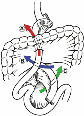 Mesenteric defects: A) transverse mesocolic; B) Petersen’s space; and C) jejunojejunostomy mesentery
- Needs to go to OT as can decompress biliary limb AND increased risk of vascular compromise
- Risk lower in laparoscopic cases and if mesenteric defects closed
- But can never prevent 100% risks
Gastric Remnant Distension (BP Limb Obstruction)
- Aetiology
- Ileus
- Mechanical obstruction
- Iatrogenic vagal injury causing decreased gastric emptying
- Gastric remnant distends and becomes necrotic
- Pt becomes hypovolaemic
- Stomach ruptures
- Massive fluid loss as well as massive 3rd spacing will cause sudden cardiac arrest
- Management
- Needs urgent decompression
- Surgical or percutaneous gastrostomy

Alimentary Limb Obstruction
- Occurs in situations like over-closure of mesenteric defects
- Causes food build up at gastro-jej, in pouch, and oesophagus
- Manage by NG decompression
- Not as severe as BP limb obstruction
Gastro-Gastric Fistula
- Rare complication of RYGB
- Can lead to
- Weight regain
- Marginal ulceration
- Aetiology
- Anastomotic leak
- Incomplete gastric division
- Marginal ulcers
- Distal obstruction
- Erosion of foreign body
- Endoscopy is diagnostic but therapeutic efficacy is very low
- Can also use PO contrast CT
- Surgery, incl. remnant gastrectomy, is the definitive treatment
- Treatment results in resolution of symptoms and improved weight loss


Marginal Ulcer
- 2-10%
- Incidence can be reduced by pre-op treatment for H. pylori
- Aetiology
- H. pylori, Smoking, NSAIDs, EtOH
- Also consider
- Poor tissue perfusion/tension
- Foreign body
- Gastro-gastric fistula
- Presentation
- Usually constant boring epigastric pain
- Nausea, GI bleed, stricture, perforation
- Usually settle with medical treatment
- Manage with high dose PPI and re-endoscopy
- Eradicate H. pylori
- Persistent ulcers may require surgical revision
- Resect ulcer/fistula and revise gastro-jej anastomosis
- Consider other causes (Gastrin levels etc.) if recurs after this
- May need reversal or gastric remnant resection
Gallstones
- Expected complication of weight loss
- 10% have symptomatic stones at 1 yr
- Can mitigate with the use of ursodeoxycholic acid
- Ursodiol 300mg po BD
- Drops risk to 2%
- But expensive
- If choledocholithiasis:
- Lap/open CBDE
- Lap/open assisted ERCP
- Enteroscopy assisted ERCP
Dumping Syndrome
- Recommend small frequent meals
- Low CHO intake
- Octreotide can help if dietary modification fails
Early Dumping Syndrome
- Occurs within 20-30mins after meal
- More common than late dumping
- More GI and less cardiovascular effects
Late Dumping Syndrome
Reflux Post Sleeve
- 20% have GORD at 5 years
- Barrett’s 10% at 10 yrs
- 2.5-5% require surgery for reflux at 5 years
- Options
- First line is anti-reflux medical therapy
- Hiatus hernia repair (with falciform sling) if hernia present and no weight regain
- Rule out and treat strictures
- RYGB if weight regain, no HH, or failure of above
Gastric volvulus
- Due to loss of fixation along greater curvature
- Symptoms
- Borchardt’s triad
- Epigastric pain
- Retching
- Inability to pass NGT
- Borchardt’s triad
- Management
- Reduce
- Operative fixation to omentum
Portal Vein Thrombosis
- Rare
- Present with pain
- Often missed on CT, need to specifically look for this
- Easily treated, but major if missed