Section: Abdominal wall and retroperitoneum Sub-section: Hernias Curriculum: Curriculum, page 2
Incidence
- Prevalence of 3% in men
- 15 per 1000 population
- M:F = 25:1
- Indirect: direct in men is 2:1
- Direct hernias in women are rare
- Femoral hernias are rare in men and uncommon in women (inguinal hernias are more common in women than femoral)
- 10% of women and 50% of men with a femoral hernia have, or will develop, an inguinal hernia
- Indirect and femoral hernias are 2 x as common on the right
- Slower descent of the testis on the Right
- Delayed obliteration of the process vaginalis
- Presence of the sigmoid colon on the left blocking the femoral canal
Aetiology
- Congenital or acquired
- Congenital
- Lack of obliteration of processus vaginalis after testicular descent
- Risks
- Prematurity
- Low birth weight
- Collagen deficiency
- Pelvic deformity
- Acquired
- Wear and tear, straining
- Abnormal fibroblastic proliferation
- Cigarette smoking
- Advanced age
- ? Malnutrition and vitamin deficiency
- Congenital
Classification
- Nyhus Classification 1991
- Type I
- Indirect hernia associated with a normal internal ring
- Type II
- Indirect hernia with an enlarged deep ring
- Inferior epigastric vessels not displaced
- The posterior wall of the inguinal canal is normal in types I & II
- Type III - Posterior wall defect
- IIIA - Direct
- IIIB - Indirect with a dilated internal ring encroaching onto the posterior wall of the inguinal canal
- IIIC Femoral
- Type I
Clinical
History
- History of lump, when, where, reducible or not?
- Key is occupation and how it affects them
- R/O weight loss or malignancy
- Previous surgery
Exam
- Inguinal
- Hernia above and medial to the pubic tubercle
- C.f. Femoral – below and lateral
- Differentiate from scrotal lesion
- Can’t get above it
- Reduces on palpation
- Positive couch impulse
- +/- Bowel sounds on auscultation
- Does not transilluminate
Management
Indications for surgery
- Primary management of inguinal hernia is surgical repair if there are no local or systemic contradictory conditions
- If surgery contra-indicated, trusses may be useful
- If asymptomatic could consider a watch and wait approach for inguinal hernias (not femoral!)
- RCT showed
- 10% will become symptomatic per year
- 40% will have been repair by 3 years and 70% by 10 years
- <0.1% risk of strangulation per year
- No increase in chronic pain for surgery vs watchful waiting
- RCT showed
- Repair types
- Open
- Lichenstein tension free mesh repair
- Various methods of suture repair
- Herniotomy (see childhood inguinal hernia)
- Laparoscopic
- TAPP
- TEP
Lichenstein Hernia Repair
- Rationale
- Inguinal hernias are caused by a metabolic disorder, which leads to a progressive destruction of the fibroconnective tissue of the groin, making the tissue unsuitable for being used in hernia repair
- Traditional tissue repairs are associated with undue tension at the suture line, which leads to more post-op pain, longer recovery time and higher recurrence rate
Technique
- A 5 cm to 6 cm transverse incision is made within a Langer’s line, beginning from the pubic tubercle 2 fingerbreadth above inguinal ligament
- External oblique aponeurosis opened in the line of its fibres
- Spermatic cord with its cremasteric covering, external spermatic vessels, and the genital nerve are freed from the inguinal floor and lifted
- Spermatic cord is also dissected free from the pubic bone area for approx 2 cm medial to the pubic tubercle to make room for extending the mesh beyond the pubic tubercle
- External oblique aponeurosis is dissected from the underlying internal oblique muscle and aponeurosis high enough to make room for 6-7cm mesh
- Sac is then dissected from the cord beyond its neck
- Inverted into the preperitoneal space (may require ligation /excision)
- If the sac is very large, the sac is divided at the midpoint of the inguinal canal.
- The proximal end is closed, dissected away from the cord structures, and inverted into the preperitoneal space
- Direct hernia bulge is large, it is inverted utilizing a purse-string on the transversalis fascia
- This is only to make the floor of the inguinal canal flat in order to facilitate placement of the mesh
- First anchoring suture of the mesh fixes the mesh to the anterior rectus sheath where it inserts into the pubic bone
- Lower edge of the mesh is sutured to the inguinal ligament in a continuous fashion - ends at the lateral border of the deep ring
- Upper tail is then crossed over the lower one and are sutured together and down onto inguinal ligament , tucked under the external oblique aponeurosis
- Upper edge of the mesh is fixed in place, care is taken to keep the mesh slightly relaxed
Advantages and Disadvantages
- Post-op pain minimal
- Recurrence rate consistently < 1%
- Can be performed under local anaesthesia
- Easy to learn
- Economical
- Disadvantages
- ?infection rate
- No clear evidence of increased infection rate
- BJS 1999 reviewed infected mesh-
- All had to be removed but rate was 1 in 1000
- ?infection rate
Suture Inguinal hernia repair
- Method
- Bassini
- Shouldice
- Division and double breasting of transversalis fascia
- Then suturing conjoint tendon to inguinal ligament with continuous suture
- Marcy
- Simple deep ring closure
Advantages and Disadvantages
- Advantages
- Can be performed under local anaesthesia
- So Age is never a factor
- Minimal equipment necessary
- Most cost-effective repair
- Incidence of dysejaculation and inguinodynia seen less frequently with pure tissue repairs
- Disadvantages
- Inadequate in the treatment of femoral hernias
- Basic pathology of herniation is not addressed
- Higher incidence of testicular atrophy - more skeletonisation of cord
- 1% incidence of chronic pain lasting beyond a year after surgery
- Cannot be relied upon for recurrent inguinal hernias
Bassini Repair
- The Bassini technique for inguinal hernia repair involves suturing the transversalis fascia and the conjoined tendon to the inguinal ligament behind the spermatic cord with monofilament nonabsorbable suture.
- It also involves the so-called Tanner slide, which is a vertical relaxing incision in the anterior rectus sheath intended to prevent tension.
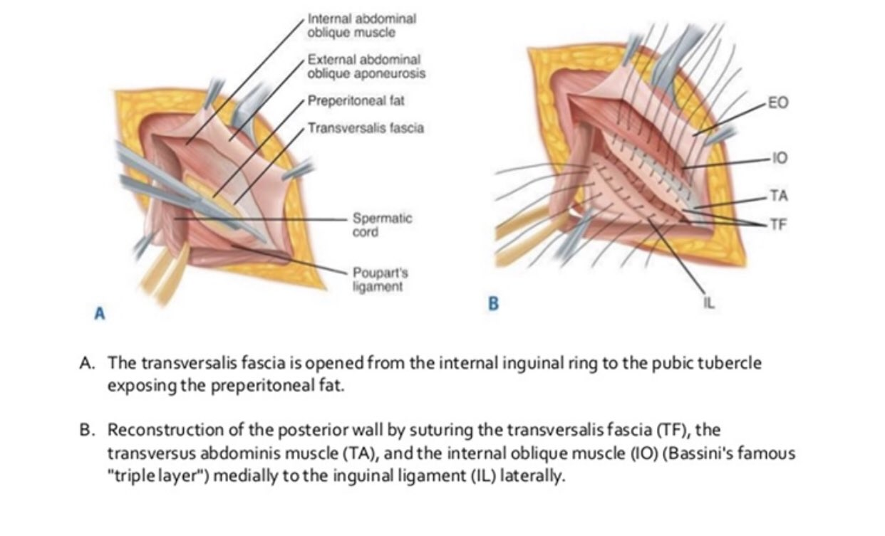
Shouldice repair
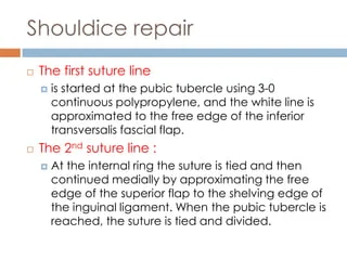
- Four-layer inguinal hernia repair performed with the patient under local anesthesia.
- Transversalis fascia is incised from the internal ring laterally to the pubic tubercle medially, and upper and lower flaps are created. These flaps are then overlapped (double-breasted) with two layers of sutures.
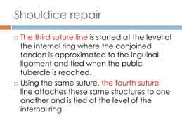
- The conjoined tendon is then sutured to the inguinal ligament, again in two overlapping layers.
- This reinforces the posterior wall and narrows the deep inguinal ring.
- The Shouldice repair is classically done with a continuous suture of 32- to 34-gauge stainless steel wire, but synthetic monofilaments (eg, polypropylene) can also be used.
- The external oblique aponeurosis is then closed in a double-breasted fashion in front of the spermatic cord.
Laparoscopic inguinal hernia repair
TAPP Laparoscopic Repair
- Trans-abdominal pre-peritoneal patch repair
- Incise peritoneum only and create a flap to do repair, and then close peritoneum over this again
- Pre-peritoneal space is then entered by incising the peritoneum transversely from the region of the median umbilical ligament laterally
- Anterior to the hernial defect
- Peritoneal flaps then developed
- Direct and small indirect sacs fully reduced
- Larger indirect sacs are part dissected and having freed the cord structures posteriorly, circumcised
- 15 x 10 cm mesh fashioned and inserted
- Medial border of mesh adjacent to symphysis pubis
- Posterior part placed well behind internal ring
- Stapled in place
- Staples applied to the pubic bone and Cooper’s ligament
- Further staples placed into muscle layers anteriorly
- None into or posterior to the iliopubic tract
- Peritoneum then reconstituted (running suture)
TEP Laparoscopic Repair
- Total Extra-Peritoneal Repair
- Good for – Recurrent, bilateral
- Becoming more common for primary hernias
- Earlier return to work with less pain
- But ? More complications and recurrence
- Contraindicated if:
- Hernia still irreducible once anaesthetised
- Previous complicated pelvic surgery
- Significant cardiovascular disease or severe COPD
Anatomy
- Space created in pre-peritoneal plane
- Safe places to staple
- Pubic tubercle
- Cooper’s ligament
- Above Iliopubic tract laterally
Technique
- Entry
- Sub-umbilical incision
- Dissect onto rectus sheath
- Mobilise laterally opposite to hernia side
- Vertical incision over muscle 2cm long ≈ 2cm from midline
- Mobilise rectus laterally
- Finger dissection into preperitoneal space
- Can usually reach down to symphysis
- Insert Hasson - inflate (or insert inflation balloon to create space)
- 20x inflate under vision, wait with tamponade, then release
- Insert 2x 5mm ports in midline
- Disection
- Identify & preserve inferior epigastrics, keep on superficial aspect
- Blunt dissection to reveal & identify
- Symphysis pubis
- Develop retroprostatic space
- ASIS
- Inguinal ligament
- Identify position of iliac vessels
- Identify deep ring
- Direct hernia will often reduce spontaneously
- Indirect
- Tease out from cord
- Careful of perforation of peritoneum
- Unequivocal identification of vas & vessels — Reduce sac fully or divide
- Mesh
- Roll 10x15cm mesh on grasper, (cut 1 corner) & insert through 10mm port
- Roll out, use cut corner for orientation
- Staple to pubic bone (above pubic bone not directly into), Cooper’s ligament, 2cm above Iliopubic tract & laterally
- Keep sac on peritoneal side, vessels on wall side
- Deflate under vision - Keep mesh in position
- Remove ports
- Close with 1 vicryl UR6 to sheath & 3-0 monocryl to skin
Complications of Hernia Repair
- Wound
- Early
- Seroma, haematoma, wound/mesh infection
- Late
- Numbness, keloid
- Early
- Testicular
- Scrotal oedema
- Impotency
- Ischaemic Orchitis
- Testicular atrophy later, esp. if divide cremasteric and don’t hitch up distal portion
- Vas injury
- Skin
- Paraesthesia - hard to fix
- Reaction to prep/tape
- Nerve entrapment
- Neuroma/Chronic pain
- Open
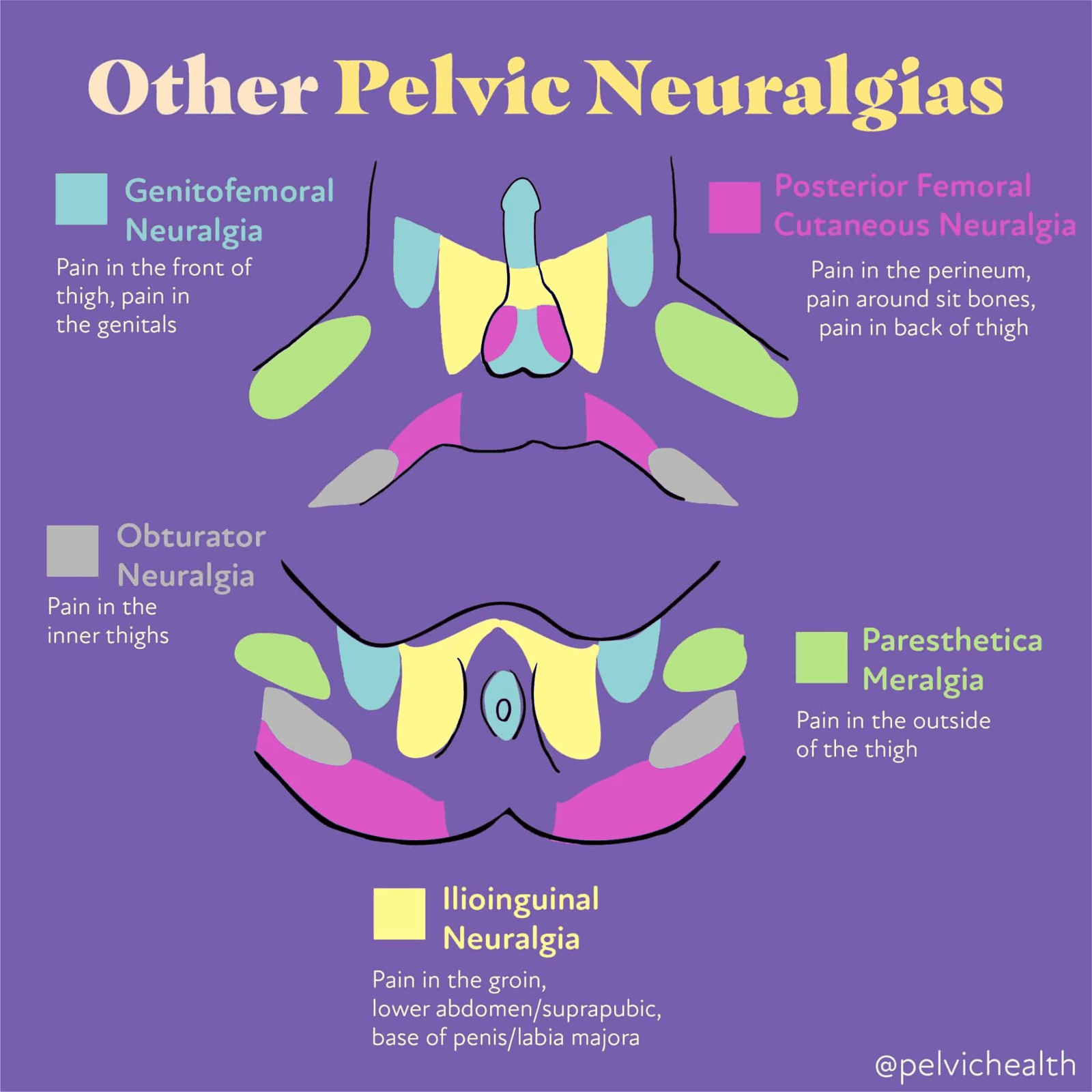
- Ilioinguinal/Iliohypogastric
- Lap
- Lat Fem Cut. Nerve
- Meralgia Parasthetica/genitofemoral
- Bowel/bladder injury
- Urinary retention/UTI
- Recurrence
Post-Hernia Repair Groin Pain
- Iliohypogastric Nerve (T12, L1)
- First branch off lumbar plexus
- Arises lateral border of psoas, travels behind kidney, then pierces fascia to reach neurovascular plane
- Pierces external oblique aponeurosis 2.5cm above superficial ring
- To suprapubic region, skin over lower rectus and mons
- Also supplies Internal Oblique (damage can precipitate hernia)
- Ilioinguinal Nerve (L1) – Collateral branch of Iliohypogastric
- Second branch off lumbar plexus, lateral border of psoas
- Pierces lower border of internal oblique and enters inguinal canal from the side (Not through Deep Ring!)
- Male
- Sensation to skin over root of penis and anterior 1/3 of scrotum, and upper medial thigh
- Muscular branches to Internal Oblique and Transverse Abdominis – Strengthen conjoint tendon
- Female
- Skin covering Mons Pubis and Labia Majora
- Lateral Femoral Cutaneous Nerve (L2, L3)
- Third branch off lumbar plexus, Lateral border of psoas, on iliacus
- Passes through inguinal ligament to anterior thigh
- Supplies parietal peritoneum of iliac fossa and skin of the lateral thigh
- Meralgia Parasthetica – pain and altered sensation of lateral thigh due to compression of Lateral Femoral Cutaneous Nerve
- Genitofemoral Nerve (Genital branch) (L1, L2)
- Arises anterior to psoas (deep to psoas fascia)
- Supplies motor to cremaster and scrotal skin
- Management (> 6 weeks = Chronic pain)
- Neuropathic meds (Amitriptyline, Gabapentin)
- Nerve block - may break the cycle of pain
- Surgery – Neurotomy +/- Remove old mesh and replace with new mesh
Recurrence
- Risk Factors for Recurrence
- Age (older age)
- Duration of hernia (short duration)
- Type of hernia (Femoral more than Inguinal)
- Coexisting medical illness
- In children, the risk factors were
- Age (very young)
- Gender (male)
- Short duration of hernia
- Side (right side)
Lap vs Open
Laparoscopic • Pros o Smaller scars o Less hematoma, seroma, wound infection o Shorter hospital stay o Faster return to activities by one week o Less chronic pain (half from 7%) – possibly even better since using absorbable tacking devices o Less numbness – ie: less ilioinguinal & iliohypogastric nerve injury o Less risk of ischemic orchitis o Can inspect contralateral side and identify hernia defects and fix at the same operation (controversial) • Cons o Not all surgeons do them o Requires GA o Longer operation by 15 minutes o Increased incidence of vascular or visceral injury (studies for this were looking at TAPP & TEP – visceral injury should be rare in TEP unless peritoneum breached) o Port site hernia – very few cases reported