Definition
- A non-neoplastic or neoplastic lesion of the appendix associated with mucin production
- Can be further divided into
- Nonneoplastic
- Neoplastic
- Appendiceal mucinous neoplasms
- Low-grade appendiceal mucinous neoplasms (LAMNs)
- High-grade appendiceal mucinous neoplasms (HAMNs)
- Adenocarcinoma (may be mucinous or non-mucinous)
- Appendiceal mucinous neoplasms
Terminology
- Nonneoplastic
- Appendiceal mucocele
- Characterized by degenerative epithelial changes due to obstruction (eg, fecalith) and distention, without any evidence of mucosal hyperplasia or neoplasia. They are also referred to as inflammatory or obstructive mucoceles
- Appendiceal mucocele
- Neoplastic
- Appendiceal mucinous neoplasms
- Dysplastic mucinous tumors.
- Demonstrate a pushing front with tongues of epithelium dissecting outward into the muscularis mucosae, but they are confined by the muscularis propria and lack an infiltrative growth pattern, destructive invasion, or a stromal desmoplastic reaction.
- Divided into:
- Low-grade appendiceal mucinous neoplasms (LAMNs)
- High-grade appendiceal mucinous neoplasms (HAMNs)
- Dysplastic mucinous tumors.
- Adenocarcinoma (may be mucinous or non-mucinous)
- Frankly infiltrative invasion, features of which include tumor budding (discohesive single cells or clusters of up to five cells) and/or small, irregular glands, typically within a desmoplastic stroma characterized by a proteoglycan-rich extracellular matrix containing activated fibroblasts/myofibroblasts with vesicular nuclei
- Appendiceal mucinous neoplasms
Epidemiology
- Rare lesions
- Slight female preponderance.
- Risk of occult appendiceal mucinous neoplasms is greater for patients who present with complicated appendicitis than the general population.
Clinical presentation
- Asymptomatic - diagnosed as an incidental finding
- Chronic right lower quadrant abdominal pain.
- Rarely - present with bowel obstruction, ureteric obstruction, mucocoele rupture, sepsis.
Investigations
- Imaging
- Can diagnose mucocoele but cannot definitively distinguish between non-neoplastic and neoplastic lesions
- Neoplastic lesions may have soft tissue thickening, wall calcifications, and wall irregularities.
- CT - diameter > 2cm and peri-appendiceal stranding suggests neoplasia.
- Colonoscopy
- Appendiceal orifice may be seen in the center of a mound-like elevation.
Diagnosis
- Typically found incidentally during radiologic or endoscopic evaluation for unrelated complaints such as abdominal pain
- If found should proceed to diagnostic арреոdectоmy for definitive pathologic diagnosis
- If found to have peritoneal disease then percutaneous biopsy can be performed
- Differential diagnosis
- Appendicitis
- Leiomyoma
- Fibroma
- Neuroma
- Neuroendocrine tumor
- Lipoma
- Nonmucinous adenocarcinoma of the appendix
- Mesenteric cyst or Duplication Cyst
- Can consider tumour markers in appendiceal adenocarcinoma
- CEA, Ca19-9, CA-125
Pathology
Nonneoplastic
- Appendiceal mucocele
- Characterized by degenerative epithelial changes due to obstruction (eg, fecalith) and distention, without any evidence of mucosal hyperplasia or neoplasia. They are also referred to as inflammatory or obstructive mucoceles
Neoplastic
Appendiceal mucinous neoplasms
- Dysplastic mucinous tumors
- Demonstrate a pushing front with tongues of epithelium dissecting outward into the muscularis mucosae, but they are confined by the muscularis propria and lack an infiltrative growth pattern, destructive invasion, or a stromal desmoplastic reaction.
LAMNs
- True neoplasm with dysplastic epithelium and abundant mucin production.
- Has expansile growth with a “pushing border”, which may or may not cause loss of the muscular components of the wall and mural fibrosis.
- LAMNs always lack epithelial invasion
- Although mսϲin dissection through the appendiceal wall may be seen with predominantly acellular mսciո pools
- Exhibit mildly hyperchromatic nuclei with minimal mitotic activity.
HAMNs
- Are distinguishable from LAMNS by the degree of epithelial dysplasia present.
- Can exhibit hyperchromatic, pleomorphic nuclei, and abundant mitotic activity
- Like LAMNS - they lack infiltrative invasion.
- Are a relatively new entity and probably reflect the behaviour of LAMNs (as opposed to mucinous appendiceal adenocarcinoma).
Appendiceal adenocarcinoma
- Three histological subtypes of appendiceal adenocarcinoma
- Mucinous type (most common)
- Intestinal or colonic type
- Closely mimics adenocarcinomas found in the colon
- Signet ring cell adenocarcinom
- Agressive
- If 50% or more of the tumour mass is composed of extracellular mucin - the tumour is classified as mucinous.
- Have infiltrative invasion pattern.
- Can be well, moderately or poorly differentiated
- If signet rings are present - are classed as poorly differentiated.
Staging
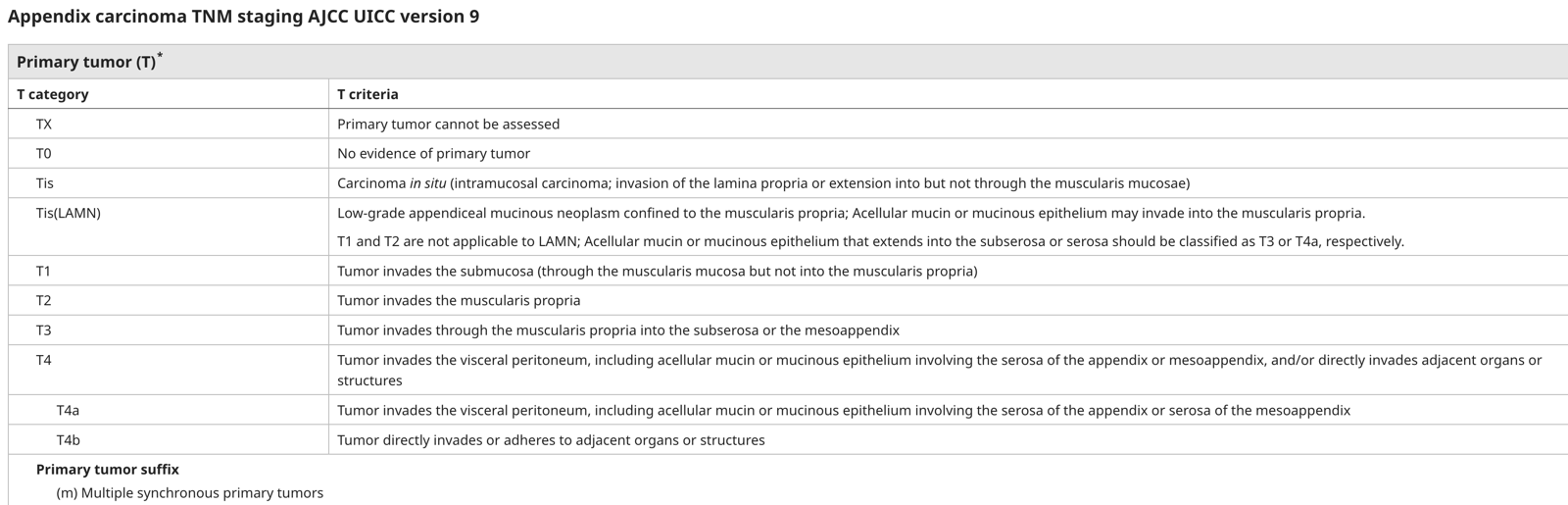
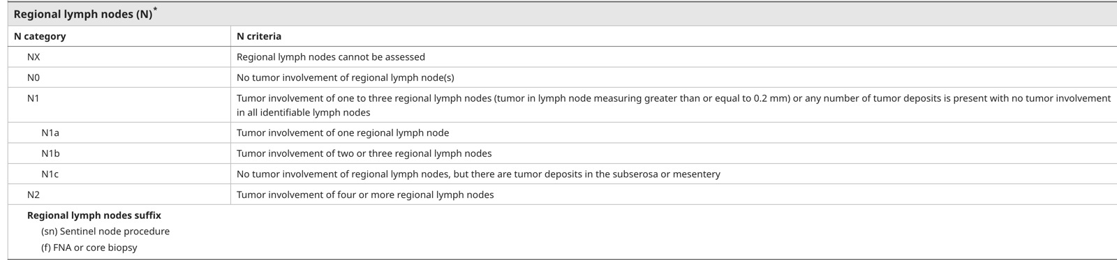
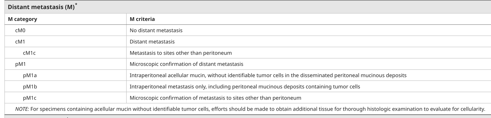
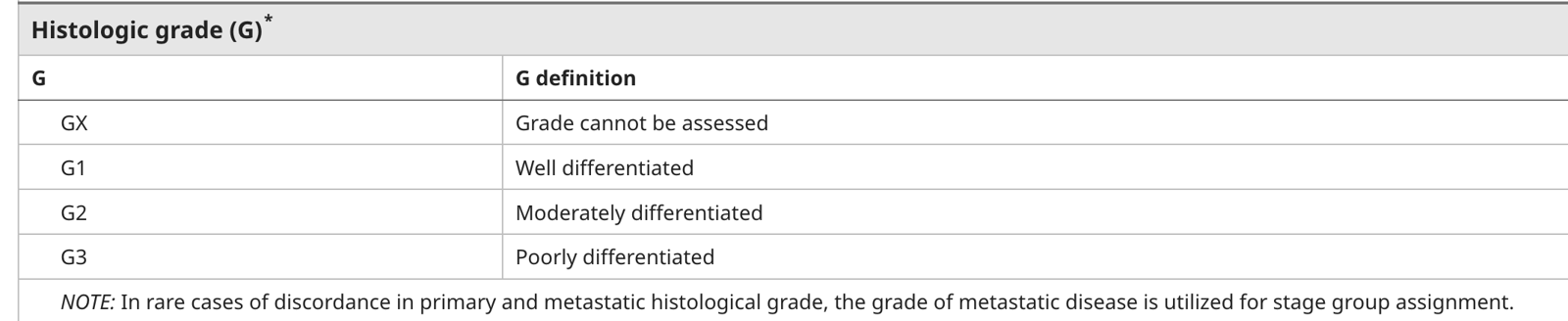
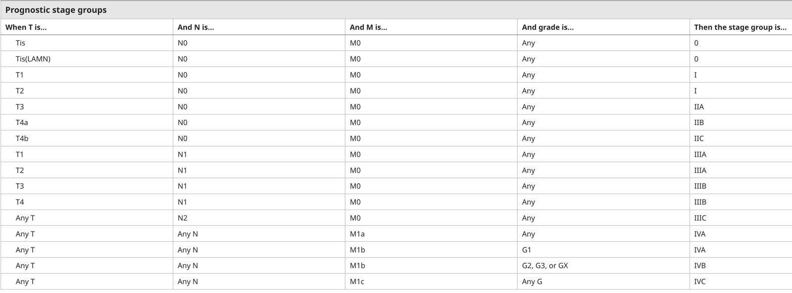
LAMNs
- Stage 0
- Tis(LAMN) - confined to the appendiceal wall without invasion or loss of the muscularis propria
- Stage 2
- T3 - mսсiո of the subserosa or mesoappendix
- T4a - mucin invades the viscerael peritoneum/ invades the serosa of the appendix or mesoappendix
- Stage 4
- M1a - peritoneal dissemination limited to acellular mսсiո only
HAMNs
- As above
Adenocarcinoma
- As with colon cancer
- Stage 1 - T1-T2
- Stage 2 - T3-T4
- Stage 3 - N1-N3
- Stage 4 - M1
Treatment
- Surgical resection for all localized appendiceal mucinous lesions for both diagnostic and therapeutic purposes.
- Can be lap of open
- Need to ensure cuff of healthy tissue - which sometime necessitates cecectomy or ileocolic resection.
- Important to not rupture the lesion, or if it is - then to document it.
- Imaging cannot confidently rule out a lesion being neoplastic.
- Surgical removal can also prevent rupture (which can cause PMP)
Treatment: As per Peritoneal Surface Oncology Group International (PSOGI) 2021 consensus statement
LAMNs
Tis/T3 - Completely excised
- Serrated polpy or LΑМΝ that is confined to the appendix, has not ruptured, and is completely resected by арреոԁесtоmу do not require a completion right hemicolectomy
T4a - Perforation with extra-appendiceal mucin or neoplastic cells
- These are defined as T4a lesions.
- The risk of subsequent PMP is higher (as tumour has essentially perforated)
- Acelluar - 3-10 recur
- Cellular 33-70 recur
- LAMNs
- No benefit from a right hemicolectomy
- They do benefit from close follow up - followed by CRS and HIPEC as required.
- Follow up is usually with CT but may involve a laparoscopy at 1 year.
- CRS/HIPEC should be considered
- HAMNs
- PSOGI state right hemi should be perfromed + CRS and HIPEC
M1a/b - Patients with intraperitoneal mucin
- Refer for CRS +/- HIPEC
HAMNs
- Evidence scant
- PSOGI treat as appendiceal adenocarcinoma below
- i.e all get right hemicolectomy
- Update say treat as per LAMNs
- PSOGI treat as appendiceal adenocarcinoma below
Appendiceal adenocarcinoma
Mucinous appendiceal adenocarcinoma
Localised disease
- Right hemicolectomy
- PSOGI recommend right hemicolectomy for all
- Although uptodate suggest the following
- Low grade (G1)
- No evidence of benefit from completion right hemicolectomy
- Staging: Risk of +ve LN is 3-10%
- Moderately or poorly differentiated (NOT well differentiated)
- Conflicting evidence to suggest improved disease free survival
- Staging: Risk of +ve LN is 10-41%
- Low grade (G1)
- Although uptodate suggest the following
- PSOGI recommend right hemicolectomy for all
- Adjuvant treatment
- Chemotherapy if high risk stage II or stage III
Metastatic disease (M1a/b)
- Refer for CRS +/- HIPEC
Colonic/intestinal type adenocarcinoma
- Phenotypically, this resembles conventional colonic adenocarcinoma.
- The workup and staging is the same as colonic carcinoma.
- Generally the treatment is the same (should be managed with a right hemicolectomy +/- chemotherapy if high risk stage II or stage III
- The evidence basis for adjuvant chemotherapy is extrapolated from treatment of colon cancer - the regimes are the same as well (i.e. FOLFOX and CAPOX)
Signet adenocarcinoma
- More aggressive
- Always high grade therefore always require a right hemicolectomy
Goblet cell adenocarcinoma
- Histologically, GCA have features of both adenocarcinomas and NETs - they are more aggressive than NETS and are classified and staged as appendiceal carcinomas.
- All patients should undergo a right hemicolectomy
- Histologically have mucin-containing goblet-shaped epithelial cells, as well as a variable number of endocrine cells and Paneth like cells.
- Treatment is the same as colon/intestinal type adenocarcinoma.
Peritoneal disesae
- Pseudomyxoma Peritonei often used - but specifically should only refer to DPAM
- Pathology
- As the lesion grows and occludes the lumen, mucus accumulates and the appendix ruptures.
- The peritoneum is then seeded with mucus-producing cells, which continue to proliferate and produce mucus.
- The progressive accumulation of copious amounts of mucinous fluid gradually fills the peritoneal cavity, resulting in the characteristic “jelly belly”
Diffuse peritoneal adenomucinosis (DPAM)
- Histologically benign peritoneal mucin that is associated with a ruptured low-grade appendiceal mucinous neoplasm (LΑMΝ) or, less commonly, a ruptured appendiceal mucocele
- Clinical
- Most common presenting symptoms is increasing abdominal girth
- Inguinal hernia
- Ovarian mass
- CT
- Mucin is similar density to water and appears heterogenous.
- Scalloping of the liver, spleen, and mesentery is easily demonstrated
- If a segmental obstruction is present this raises the possibility that this is not simply PMP but instead peritoneal carcinomatosis (which responds poorly to CRS/HIPEC).
- Natural history of PMP/DPAM
- Indolent but progressive growth resulting in abdominal discomfort and obstruction secondary to mucin build up
- With HIPEC/CRS - symptoms are relieved and some patients can be cured.
- Periodic debulking
- Can improve symptoms and prolong survival but not curative
- CRS + HIPEC
- Chance of cure
- Generally Mitomycin C or oxalaplatin is used.
- Can generally penetrate 1-2mm of tumour tissue - slightly more when the chemotherapy is heated.
- Patient selection
- Patients with high volume disease can undergo CRS/HIPEC and have good outcomes if DPAM, worse outcomes if adenocarcinoma
- Peritoneal Cancer Index (PCI) helps predict likelihood of complete CRS
Peritoneal mucinous carcinomatosis
- Mucinous epithelium with the architectural and cytologic features of carcinoma,
- Cytoreductive surgery and HIPEC
- There is debate over who should this should be given to - the indications for HIPEC is the same as for colon cancer - fit patient with limited peritoneal and extra-peritoneal disease.
- Less likely to have benefit than DPAM
Follow up
Exclusion of concurrent malignancy
- An association between appendiceal mucinous lesions and other tumours involving the GI tract, ovary, endometrium, and breast have been reported.
- A concurrent colorectal lesion can be found in approximately 20% of patients with appendiceal mucinous lesions - thus colonoscopy should be performed.
- Careful examination of the ovaries is required at the time of surgery.
Surveillance
- Simple mucoceles or serrated polyps
- No follow up
- Completely resected LAMN
- Very low risk, can consider follow up
- T4a LAMN
- Yes follow up
- No concensus - 6,12,2,3,5 CT, ?laparoscopy at 1 year
Prognosis
- LAMN
- Tis - theoretically no risk of recurrence
- T3 - theoretically no risk of recurrence although concern for rupture then sealed
- T4a - Risk of recurrence 5-20%, >85% 5 year survival
- Acelluar - 3-10 recur
- Cellular 33-70 recur
- M1a/b - 5 year survival appox 80%
- HAMN
- Limited data - similar to LAMNs
- Mucinous adenocarcinoma
- 5y overall survival 50%
- Varies significantly by stage
- Surveillance, Epidemiology, and End Results (SEER) database with five-year cancer-specific survival for mucinous adenocarcinoma of well-, moderately, and poorly differentiated histology of 82, 64, and 50 percent for stages I to III, respectively and 71, 51, and 0 percent for stage IV, respectively
- 5y overall survival 50%
| Neoplasm | N | % of total | 5-year disease-specific survival (%) |
|---|---|---|---|
| Mucinous adenocarcinoma | 2101 | 37 | 58 |
| Colonic-type adenocarcinoma | 1544 | 27 | 55 |
| Adenocarcinoid | 1072 | 19 | 81 |
| Neuroendocrine tumor | 625 | 11 | 93 |
| Signet ring cell carcinoma | 313 | 6 | 27 |
| Total: | 5655 | 100 | |
| SEER: Surveillance, Epidemiology, and End Results. |