Development
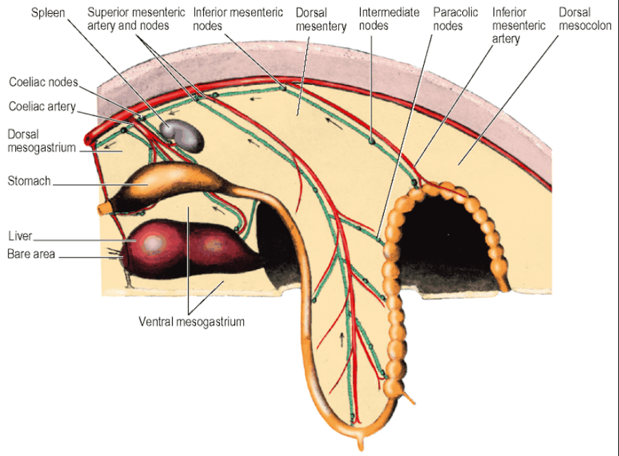
- Just before the 6th week of embryonic life:
- Alimentary canal is a simple tube
- Whole length supported by a dorsal mesentery attached in the midline in front of the Aorta
- Three gut arteries leave Aorta and pass ventrally to supply tube
- Coeliac Artery - most cranial – passes in the Dorsal Mesogastrium to supply the Foregut
- Superior Mesenteric Artery - passes through the Dorsal Mesentery to supply the Midgut
- Inferior Mesenteric Artery - passes through the Dorsal Mesocolon to supply the Hindgut
- Derivatives of Foregut, i.e. liver and pancreas, and spleen supplied with blood by the Coeliac Artery
Aorta
Course and Relations
- Origin: The continuation of the Thoracic Aorta on passing behind the Median Arcuate Ligament and between the Crura of the Diaphragm
- Anterior to body of T12 Vertebra
- Passes downwards behind the peritoneum on the bodies of the Lumbar Vertebrae
- Inclines slightly to the left
- Left Sympathetic Trunk at its left margin
- Termination: Divides on the body of L4 into two Common Iliac Arteries
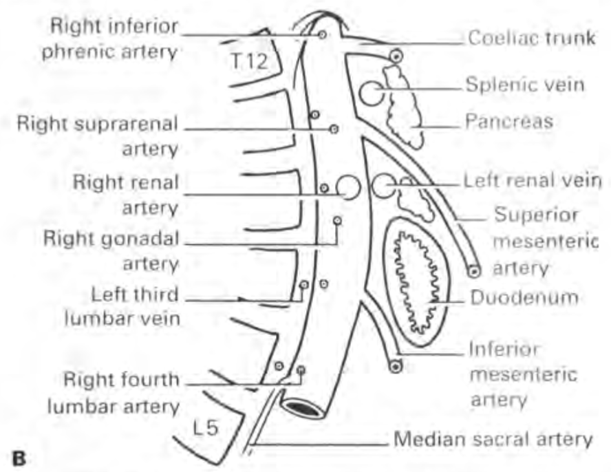
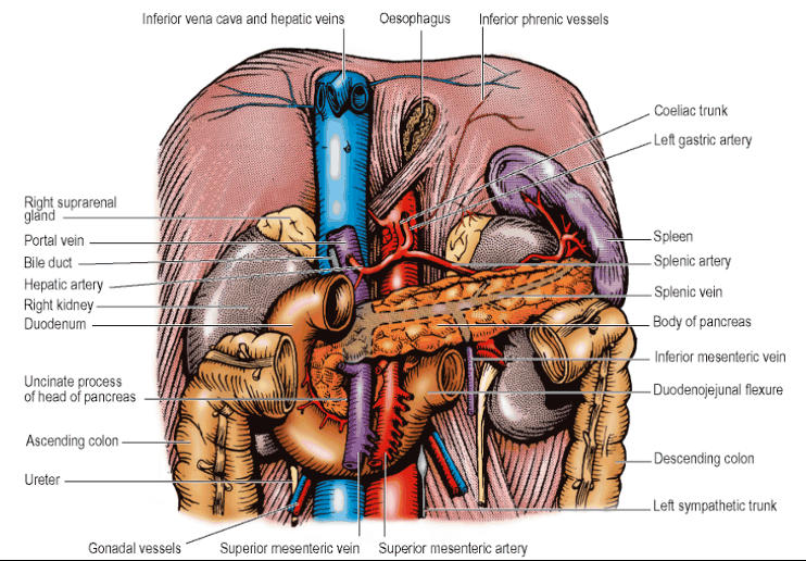
- Between the origins of the Coeliac Trunk and the Superior Mesenteric Artery, the Aorta is crossed by the Splenic Vein and Body of Pancreas
- Between the origins of the Superior and Inferior Mesenteric Arteries, the Aorta is crossed by the Left Renal Vein, the Uncinate Process of the Pancreas and the Third Part of the Duodenum
Branches
-
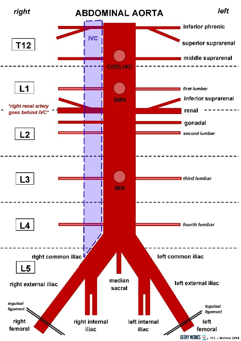
Main branches fall into 3 groups:
- Single ventral arteries to the gut and derivates
- Coeliac Artery - T12
- Superior Mesenteric Artery - L1
- Inferior Mesenteric Artery - L3
- Paired branches to other viscera
- Suprarenal Arteries - L1
- Renal Arteries - L2
- Gonadal Arteries - L2
- Paired branches to abdominal wall
- Inferior Phrenic Arteries - T12
- Lumbar Arteries - L1-L4
- Single ventral arteries to the gut and derivates
-
Median Sacral Artery - L4
- Small posterior branch
- Leaves the Aorta a little above its bifurcation
- Runs in the midline over the Sacral Promontory into the Hollow of the Sacrum
- Anastomoses with the Lateral Sacral Arteries
- Gives minute branches to the Rectum
-
Relevance of branches of the Abdominal Aorta for surgical management:
- An important weakness of endovascular repair of AAAs is back-bleeding or endoleak formation due to incomplete sealing or bridging of aortic branches ostia by endoluminal stents
Inferior Phrenic Artery
- First branches of the Abdominal Aorta T12
- May arise by a common stem just above the Coeliac Trunk
- Each slopes upwards over the Crus of the Diaphragm, which they supply
- Left Artery passes behind the Oesophagus
- Right Artery passes behind the Inferior Vena Cava
- Give off small Suprarenal branches
Suprarenal Arteries
- Arise from the Aorta between its Inferior Phrenic and Renal branches at L1
- Run laterally across the Crus of the Diaphragm
- Right Suprarenal Artery passes behind the Inferior Vena Cava
- Left Suprarenal Artery passes behind the posterior wall of the Lesser Sac
Renal Arteries
- Arise at right angles from the Aorta
- At the level of L2 Vertebra
- Left Renal Artery:
- Shorter than the Right
- Crosses the Left Crus and Psoas
- Behind the Left Renal Vein
- Both Left Renal Artery and Vein are covered by the Tail of the Pancreas and Splenic Vessels
- Right Renal Artery:
- Crosses the Right Crus and Psoas
- Behind the Inferior Vena Cava and short Right Renal Vein
- These structures separate the Right Renal Artery from the Head of the Pancreas and Bile Duct and from the 2nd part of the Duodenum
- Each Renal Artery gives small Suprarenal and Ureteric branches
- One or two Accessory Renal Arteries arise frequently from the Aorta (above or below the main artery)
Gonadal Arteries
- Similar origin and course in both sexes
- Arise near the front of the Aorta, below the Renal Arteries
- Arise well above the origin of the Inferior Mesenteric Artery
- Slope steeply downwards over Psoas and Genitofemoral Nerve
- Right Gonadal Artery
- First crosses the Inferior Vena Cava
- Right Gonadal Artery crossed by the 3rd part of the Duodenum and Root of the Mesentery
- Cross the Ureter, supplying its middle portion
- Crossed by the Colic Vessel
- Left Gonadal Artery
- Crossed by the Inferior Mesenteric Vein
- Reach Pelvic Brim about halfway between the Sacroiliac Joint and the Inguinal Ligament
- After this point, their course in each sex is different
- Testicular Artery
- Runs along the Pelvic Brim above the External Iliac Artery
- Enters the Deep Inguinal Ring
- Passes in the Spermatic Cord to the Testis
- Ovarian Artery
- Crosses the Pelvic Brim and External Iliac Vessels
- Enters the Suspensory Ligament
- Pass to the Ovary and Uterine Tube
- Testicular Artery
Subcostal Arteries
- Arise from the lowest part of the Thoracic Aorta
- Each enters the abdomen beneath the Lateral Arcuate Ligament
- Runs between the Subcostal Nerve and Vein on the anterior surface of the Lumbar Fascia over Quadratus Lumborum behind the Kidney
- Passes laterally into the neurovascular plane of the anterior abdominal wall (Between Internal Oblique and Transversus)
Lumbar Arteries
- Four in number (L1-L4)
- Leave the Abdominal Aorta opposite the bodies of the upper four vertebrae
- Pass behind the Lumbar Sympathetic Trunks and Fibrous arches in the Psoas
- On the right:
- Upper two Lumbar Arteries posterior to Right Crus of the Diaphragm
- Lower two Lumbar Arteries posterior to the Inferior Vena Cava
- On the left:
- Uppermost Lumbar Artery posterior to the Left Crus of the Diaphragm
- Each gives off posterior and spinal branches
- Pass laterally behind Psoas muscle
- Three upper arteries pass laterally behind Quadratus Lumborum into the neurovascular plane between Transversus Abdominis and Internal Oblique
- Fourth Lumbar Artery – passes across to the neurovascular plane in front of the Quadratus Lumborum, along the upper margin of the Iliolumbar Ligament
- No Fifth Lumbar Artery
- Its place is taken by the Lumbar branch of the Iliolumbar Artery (a branch of the Internal Iliac Artery)
- Ascends from the Pelvis in front of the Lumbosacral Trunk
- Passes laterally behind the Obturator Nerve and Psoas Muscle
- Supplies Psoas and Quadratus Lumborum
- Gives a spinal branch which enters the L5-S1 intervertebral foramen
- Iliac Branch runs in the Iliac Fossa, supplying Iliacus and the Ilium
- Ends in the anastomosis at the ASIS
Common Iliac Arteries
- Formed by the bifurcation of the Aorta on the body of L4
- To the left of midline
- Thus, Right Common Iliac Artery longer than the left
- Each passes to the front of the Sacroiliac Joint
- Bifurcation occurs into the External and Internal Iliac Arteries
- Ureter lies in front of this bifurcation or the very beginning of the External Iliac Artery
- Left Common Iliac Artery
- Crossed by the Inferior Mesenteric (Superior Rectal) Vessels lying beside the Ureter behind the Apex of the Mesocolon
- Each crossed by the Sympathetic contributions to the Superior hypogastric plexus
- Sympathetic trunk passes behind the Common Iliac Artery
External Iliac Artery
- Continues in the line of the Common iliac artery
- Passes along the Pelvic Brim on Psoas muscle
- Passes beneath the Inguinal Ligament
- Enters the Femoral Sheath as the Femoral Artery
- Two branches given off just above the Inguinal Ligament:
- Inferior Epigastric Artery: Ascends along the medial margin of the Deep Inguinal Ring
- Deep Circumflex Iliac Artery: Runs above the Inguinal Ligament
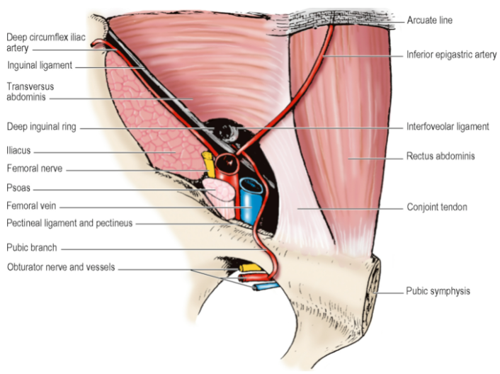
- Surface Marking of the Common Iliac and External Iliac Arteries: Along a line from the Aortic Bifurcation to the midpoint between the ASIS and Pubic Symphysis
- Common Iliac Artery corresponds to the Upper 1/3 and External Iliac Artery to the lower 2/3 of this line
- Bifurcation of the Common into the External and Internal Iliacs is 3cm from the midline, level with the Tubercles of the Iliac Crests (the Intertubercular Plane)
Blood Supply of Foregut
Arterial
- Coeliac Artery
- Origin: Front of the Abdominal Aorta between the crura of the diaphragm
- A little below the Median Arcuate Ligament
- At the level of T12 vertebra body
- Flanked by Coeliac group of Pre-Aortic Lymph Nodes
- Coeliac Ganglia lie one on each side – send sympathetic nerves carried along its branches
- Supplies: The GI tract from the lower part of the oesophagus down to the opening of the bile duct into the duodenum + Foregut derivatives (liver and pancreas) and the spleen
- Branches: Left Gastric Artery, Splenic Artery, and Common hepatic artery
- Course: Divides at the upper border of the Pancreas into three branches behind the peritoneum of the posterior wall of the Lesser Sac
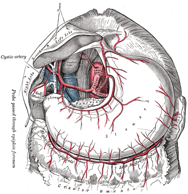
- Course:
- Runs upwards across the left crus to the oesophageal opening in the diaphragm
- Gives off oesophageal branches
- Turns antero-inferiorly raising a small fold of peritoneum, the Left Gastropancreatic Fold
- Turns right in the Lesser Omentum along the Lesser Curvature
- Supplies stomach
- Branches:
- Oesophageal branches
- Course:
- Passes to the left
- Tortuous
- Crests of its waves appear above the Pancreas
- From the middle part, may give off a Posterior Gastric Artery to the stomach, which raises a fold of the parietal peritoneum called the Gastrosplenic Ligament, as it arches forwards towards the Stomach
- Runs across the Left Crus and Left Psoas to the hilum of the Left Kidney
- Turns forward in the Splenorenal Ligament to the hilum of the Spleen
- Gives off 6 Short Gastric Arteries, which run in the Gastrosplenic Ligament
- Gives off the Left Gastroepiploic Artery – runs to the right in the Greater Omentum
- Gives branches to the Stomach and Omentum
- Breaks up into terminal splenic branches
- Branches:
- Common hepatic artery Course
- Passes over upper border of pancreas
- Then downwards and to the right behind the peritoneum of the posterior abdominal wall (in the lesser sac) as far as the first part of the duodenum
- Turns forward at the opening into the lesser sac (epiploic foramen) and turns upwards into the space between the 2 layers of the lesser omentum
- Becomes the hepatic artery
- Meets the bile duct
- Lies on left side of bile duct
- Both bile duct and hepatic artery are in front of the portal vein
- All 3 lie between duodenum and porta hepatis surrounded by peritoneum at free edge of lesser omentum
- NOTE: ‘Common hepatic’ becomes ‘hepatic proper’ after it gives of the GDA
- Meets the bile duct
Hepatic artery
- Reaches porta hepatis and divides:
- Right hepatic artery – right half of liver
- Left hepatic artery – left half of liver
Other branches
-
Right gastric
- Leaves common hepatic as it turns into lesser omentum to reach the stomach
-
Gastroduodenal
- Passes down behind first part of duodenum to the left of the portal vein
- Then divides into 2
- Right gastroepiploic artery
- Passes forward between first part of duodenum and pancreas
- Turns to the left to enter between the 2 leaves of the greater omentum at their attachment over the front of the head of the pancreas
- Superior pancreaticoduodenal artery
- Divides into 2 branches – encircle the head of the pancreas in the concavity of the duodenum down to the entrance of the bile duct
- Anterior branch (small)
- Posterior branch (large)
- Both branches anastomose with similar branches of the ‘inferior pancreaticoduodenal branch’ (from SMA)
- Bile duct entrance marks the junction of foregut/midgut – is the meeting place of the arterial distributions of the respective arteries (coeliac/SMA)
- Pancreaticoduodenal arteries supply:
- Duodenum
- Adjacent head of pancreas
- Divides into 2 branches – encircle the head of the pancreas in the concavity of the duodenum down to the entrance of the bile duct
- Right gastroepiploic artery
-
Entrance of Bile Duct marks junction between Foregut and Midgut
- Meeting place of the arterial distributions of the Coeliac and Superior Mesentery Arteries
Venous Drainage
- Reaches the Liver via the Portal Vein
- Right and Left Gastric, Right and Left Gastroepiploic and Short Gastric Veins run with corresponding arteries
- Netters page 442
- Drain the lower third of the Oesophagus in the Posterior Mediastinum
- Drains through Oesophageal opening in the Diaphragm to the Left Gastric Vein
- Oesophagus above this level drains into the Azygos system of veins
- Runs to the left along the Lesser Curvature up to the Oesophagus
- Passes medially behind the peritoneum of the posterior wall of the Lesser Sac to join the Portal Vein at the upper border of the 1st part of the Duodenum
- Runs along the Lesser Curvature to the Pylorus
- Empties into the Portal Vein
- Receives the Prepyloric Vein which ascends in front of the Pylorus
Short Gastric Veins and Left Gastroepiploic Veins
- Run with arteries through the Gastrosplenic Ligament and Greater Omentum to the Splenic Hilum
- Empty into the Splenic Vein
- Begins in Splenic Hilum
- Receives Short Gastric Veins and Left Gastroepiploic Veins
- Passes to the right with the Tail of the Pancreas, below the Splenic Artery, in the Splenorenal Ligament
- Continues to the right posterior to the Body of the Pancreas
- During its course, it lies on the hilum of the Left Kidney, the Left Psoas muscle, the Left Sympathetic Trunk, the Left Crus of the Diaphragm, the Aorta, the Superior Mesenteric Artery, and the Inferior Vena Cava
- Lies anterior to the Left Renal Vein along the upper border of that vessel
- Receives the Inferior Mesenteric Vein as it lies in front of the Left Crus of the Diaphragm
- Joins the Superior Mesenteric Vein at a right angle in front of the Inferior Vena Cava to form the Portal Vein
- Receives many tributaries from the Tail, Body, Neck and Head of the Pancreas
Right Gastroepiploic Vein
- Runs to the right in the Greater Omentum
- Descends over the front of the Pancreas
- Joins the Superior Mesenteric Vein at the lower border of the Neck of the Pancreas
Superior Pancreaticoduodenal Vein
- Ascends behind the head of the Pancreas to join the Portal Vein at the upper border of the Pancreas
- Portal vein supplies 70% of blood flow to liver
- Formed from confluence of splenic and SMV at neck of pancreas
- IMV drains at right angles into splenic vein or sometimes directly into SMV
- Portal vein receives majority of venous drainage of gastrointestinal system
- Receives left and right gastric and superior pancreaticoduodenal veins
- Most of tributaries of portal vein enter on its sides or posteriorly
- Thus space can be created bluntly between PV/SMV and neck of pancreas
- Portal vein then ascends to join porta hepatis in free margin of epiploic foramen lying behind the CBD and hepatic artery
- At porta hepatis it divides into a shorter but wider right branch and longer but narrower left branch
Blood Supply of Midgut
Arterial
- Superior Mesenteric Artery
- Supplies gut from entrance of Bile Duct to a level just short of the Splenic Flexure of the Colon
- Arises from the front of the Aorta 1cm below the Coeliac Trunk
- At level of L1 vertebra
- Course:
- Directed steeply downwards behind the Splenic Vein and Body of Pancreas
- Superior Mesenteric Vein on the right side
- Enters upper end of Small Bowel Mesentery
- Passes down to the right along the Root of the Mesentery
- NB: Terminates in the Terminal Ileum at the apex of the loop of the midgut (site of attachment of the Vitello-Intestinal Duct) – approximately 60cm proximal to the caecum
- Relations:
- Posterior: (in order from superior to inferior)
- Left Renal Vein
- Uncinate Process of the Pancreas
- 3rd Part of the Duodenum
- Posterior: (in order from superior to inferior)
- Branches:
- Inferior Pancreaticoduodenal Artery
- Jejunal Branches
- Ileal Branches
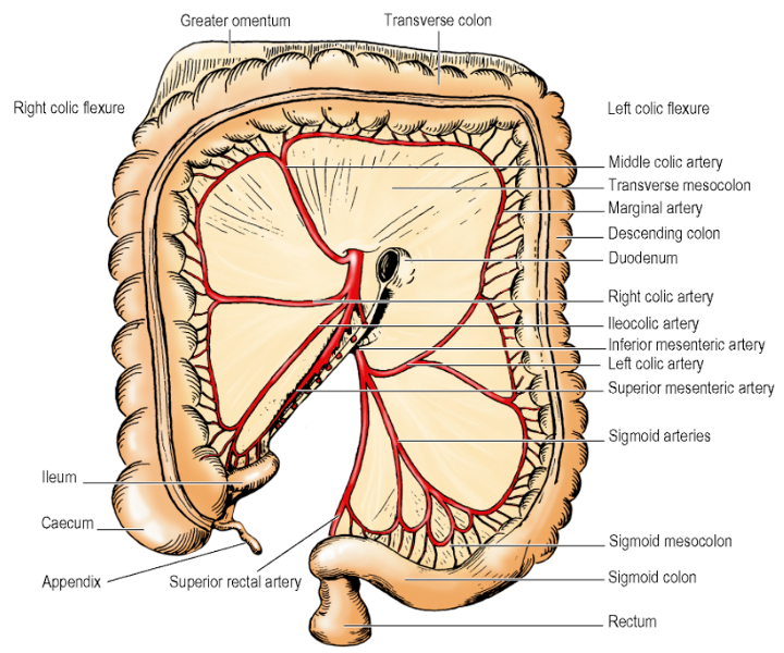
- Ileocolic Artery
- Right Colic Artery
- Middle Colic Artery
- Inferior Pancreaticoduodenal Artery
- Arises from posterior surface of Superior Mesenteric Artery
- Variation: May come off 1st Jejunal Branch
- Divides into anterior and posterior branches – run in the curve between the Duodenum and the Head of the Pancreas
- Supplies Duodenum and Head of Pancreas
- Anastomose with the terminal branches of the Superior Pancreaticoduodenal Artery
- Jejunal and Ileal Branches
- Arises from the left of the main trunk of the Superior Mesenteric Artery as it passes down between the two layers of the Mesentery
- Ileocolic Artery
- Arises from the right side of the Superior Mesenteric Artery low down in the base of the Mesentery
- Descends to the right iliac fossa and divides into superior and inferior branches
- Superior branch – Runs up along the left side of the Ascending Colon to anastomose with the Right Colic Artery
- Inferior branch – Runs to the ileocolic junction and gives off Anterior and Posterior Caecal Arteries, an Appendicular Artery, and an Ileal Branch which ascends to the left on the ileum to anastomose with the terminal branch of the Superior Mesenteric Artery
- Right Colic Artery
- Arises from the right side of the Superior Mesenteric Artery or in common with the Ileocolic Artery
- Runs across the Right Psoas Muscle, Gonadal vessels, Ureter and Genitofemoral Nerve, and Quadratus Lumborum, just behind the peritoneal floor of the Right Infracolic compartment
- Divides near the left side of the Ascending Colon into two branches
- Descending branch – runs down to anastomose with the Superior Branch of the Ileocolic Artery
- Ascending branch – runs up across the inferior pole of the Right Kidney to the Hepatic Flexure where it anastomoses with a branch of the Middle Colic Artery
- Anatomical Variation:
- Right Colic Artery present in only 10-63% of cases
- Right Colic Artery may arise from the Ileocolic Artery or the Middle Colic Artery
- Middle Colic Artery
- Arises from the right side of the Superior Mesenteric Artery, as it emerges at the lower border of the Neck of the Pancreas
- Descends between the two leaves of the Transverse Mesocolon
- Lies to the right of the midline
- At the intestinal border of the Transverse Mesocolon, it divides into right and left branches
- Right branch – anastomoses with the ascending branch of the Right Colic Artery
- Left branch – supplies the Transverse Colon almost to the Splenic Flexure where it anastomoses with a branch of the Left Colic Artery
- Anatomical Variation:
- Branches of the Middle Colic Artery mostly arise from the main trunk (64%)
- One or two arterial branches may originate directly from the Superior Mesenteric Artery in 32% and 3.3% of cases, respectively
- Multiple Middle Colic Arteries (2 or 3 branches) could arise directly from the Superior Mesenteric Artery (7.2%)
Venous Drainage
Venous Drainage of Midgut
- Each branch of the Superior Mesenteric Artery is accompanied by a vein
- All these veins flow into the Superior Mesenteric Vein
- Lies to the right of the artery
- Crosses the 3rd part of the Duodenum and Uncinate Process of the Pancreas
- Joined by the Splenic Vein behind the Neck of the Pancreas to form the Portal Vein
- Portal vein continues upwards behind the 1st part of the Duodenum
Blood Supply of the Hindgut
Arterial supply
- Supplies as far as the upper third of the Anal Canal, i.e. to the level of the Pectinate Line
- Arises from front of Aorta:
- Behind the inferior border of the 3rd part of the Duodenum
- Opposite L3 vertebra
- At the level of the umbilicus
- 3-4cm above the Aortic Bifurcation
- Smaller than the Superior Mesenteric Artery
- Runs obliquely down to the pelvic brim, immediately beneath the peritoneal floor of the left infracolic compartment
- Gives off the:
- Left Colic Artery
- Sigmoid Arteries
- Crosses the Pelvic Brim at the bifurcation of the Left Common Iliac Vessels over the Sacroiliac Joint
- Then converges towards the Ureter with the Inferior Mesenteric Vein lying in between them at the apex of the attachment of the Sigmoid Mesocolon
- Branches cross to the left in front of the Ureter
- Continues as the Superior Rectal Artery beyond the Pelvic Brim within the roof of the Sigmoid Mesocolon
- Left Colic Artery
- Passes upwards and to the left behind the peritoneum
- Divides into an ascending and a descending branch after a short course
- Ascending branch:
- Continues laterally and upwards
- Crosses the Left Psoas, Gonadal Vessels, Ureter, Genitofemoral Nerve, and Quadratus Lumborum
- Crossed anteriorly by the Inferior Mesenteric Vein
- Descending branch:
- Passes laterally and downwards
- Branches of these two arteries anastomose with each other as well as the left branch of the Middle Colic Artery and with the highest Sigmoid Artery
- Sigmoid Arteries
- 2-4 branches
- Pass between layers of the Sigmoid Mesocolon
- Last sigmoid branch anastomoses with the 1st branch of the Superior Rectal Artery
Venous Drainage
- Runs up in the root of the Sigmoid Mesocolon
- To the left of the Superior Rectal Artery to the Pelvic Brim
- Renamed the Inferior Mesenteric Vein above the Pelvic Brim
- Receives tributaries identical with the branches of the Inferior Mesenteric Artery
- Runs vertically upwards to the left of the Inferior Mesenteric Artery
- Lies beneath the peritoneal floor of the left infracolic compartment
- Lies on the Left Psoas, in front of the Gonadal vessels, Ureter, and Genitofemoral Nerve
- Lies to the left of the Duodenojejunal Flexure at the upper limit of the left infracolic compartment just below the attachment of the Transverse Mesocolon
- Curves to the right and often raises a ridge of peritoneum – the Paraduodenal recess
- Passes behind the lower border of the Body of the Pancreas
- In front of the Left Renal Vein
- Joins the Splenic Vein
- Variation: Curves to the right more sharply and…
- Passes behind the Pancreas
- Below and Parallel with the Splenic Vein
- In front of the Superior Mesenteric Artery
- Open directly into the Superior Mesenteric Vein