Pelvic Cavity
Bony Pelvis
- Overview:
- Hip bone, Sacrum, and Coccyx
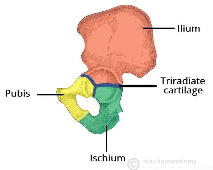
- Hip bones formed by 3 boes - ilium, ischium and pubis
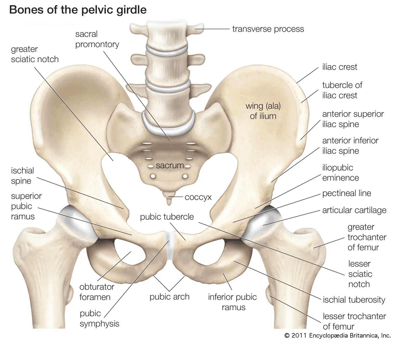
- Brim of cavity: Ala of each Ilium
- Projects up to form the Iliac Fossa, part of the posterior abdominal wall
- Pelvic Brim:
- Formed in continuity by the:
- Pubic Crest
- Pectineal Line of the Pubis
- Arcuate Line of the Ilium
- Ala and Promontory of the Sacrum
- Formed in continuity by the:
- Hip bone, Sacrum, and Coccyx
- Plane of brim is oblique – lying at 60° with the horizontal
- Vagina lies in same plane
- From Pelvic Brim, the Pelvic Cavity projects back to the buttocks
- Sex differences:
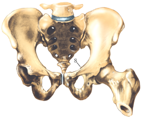
- Male pelvis makes an acute subpubic angle, pointed like a gothic arch
- Female pelvis makes a wide subpubic angle, rounded like a roman arch
- Outline of Pelvic Brim:
- Males: Sacral promontory indents outline, brim is widest towards the back, heart-shaped outline
- Females: Sacral promontory indents outline less, brim widest further forwards, transversely oval outline
- Position of Pelvis
- In erect patient, the Anterior Superior Iliac Spine (ASIS) and upper margin of the Symphysis Pubis lie in the same vertical plane
- Upper border of the Symphysis Pubis, Spine of Ischium, Tip of Coccyx, Head of Femur, and Apex of the Greater Trochanter lie in same horizontal plane
- Lower poles of the Ovaries and Seminal Vesicles also lie in this plane
- Plane passes through Pelvic Cavity at a level with the tip of the finger of the clinician during rectal or vaginal examination
Pelvic Walls
Overview
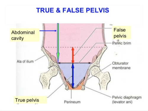
- Pelvic Brim divides the ‘False Pelvis’ (above the brim and part of the abdominal cavity) from the ‘True Pelvis’ or Pelvic Cavity (below the brim)
- Muscles of True Pelvis:
- Obturator Internus and Piriformis
- Levator Ani and Coccygeus (constitute the Pelvic Floor or Pelvic Diaphragm)
- Pelvic Side Wall:
- Formed by the hip bone clad with Obturator Internus and its Fascia
- Pelvic Posterior Wall:
- Sacrum
- Piriformis passing laterally into the Greater Sciatic Foramen
Piriformis
Origin
- Front of the middle 3 pieces of its own half of the Sacrum, Lateral Mass of Sacrum extending medially between the Anterior Sacral Foramina
- Emerging Sacral Nerves and Sacral Plexus lie on the muscle
- Runs transversely to the Greater Sciatic Foramen
- In the buttock:
- Upper border lies alongside Gluteus Medius
- Lower border lies above the Superior Gemellus
- Converges into a rounded tendon inserted into the medial surface of the upper border of the Greater Trochanter
- Pelvic surface of muscle and Sacral Plexus covered by Pelvic Fascia attached to the Sacral Periosteum at the margin of the muscle Surface Marking
- Lower border of Piriformis – midpoint of a line between the Posterior Superior Iliac Spine (indicated by an overlying dimple on the surface of the back) and the tip of the Coccyx to the Tip of the Greater Trochanter Nerve Supply
- Anterior rami of S1 and S2
Obturator Internus
Overview
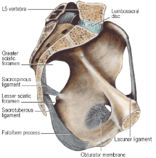
- Obturator foramen covered by the Obturator Membrane (a felted mass of fibrous tissue)
- A gap above converts the Obturator Notch into a canal for the Obturator Nerve and Vessels
- Obturator internus arises from the whole membrane and from the bony margins of the foramen
- Origin extends posteriorly as high as the Pelvic Brim and across the flat surface of the Ischium to the margin of the Greater Sciatic Notch
- Origin extends down to the Falciform Ridge on the Ischial Tuberosity
Course
- Muscle fibres converge towards the Lesser Sciatic Notch
- Tendon takes a right-angled turn to pass into the buttock
- Bone here lined by Hyaline cartilage
- Separated from the tendon by a bursa
- As the muscle emerges into the buttock, it is reinforced by the additional muscle fibres of the Superior and Inferior Gemelli muscles, arising from the margins of the Lesser Sciatic Notch
- Tendon inserts into the medial surface of the Greater Trochanter above the Trochanteric Fossa
Nerve Supply
- Own nerve (L5, S1, S2)
Obturator Fascia
- Covers Obturator Internus
- Attached to bone at the margins of the muscle and fuses below with the Falciform Process of the Sacrotuberous Ligament on the Ischial Tuberosity
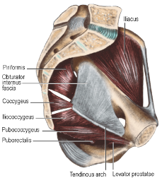
- Tendinous arch of the origin of Levator Ani slopes across the Obturator Internus Fascia
- Pelvic Cavity lies above the line
- Ischioanal Fossa below the line
Pelvic Floor
Overview
- Consists of the Pelvic Diaphragm slung around the midline viscera (Urethra, Anal Canal, and Vagina)
- Muscles of Pelvic Floor: Levator Ani and Coccygeus
- Arise in continuity from the Pubic Body, Tendinous Arch over the Obturator Fascia and Ischial Spine
- Insert into the Coccyx and Postanal Plate (a.k.a. Anococcygeal Ligament)
- From origin, muscles sweep downwards and backwards to the midline
Levator Ani
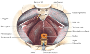
- Consists of two main parts: Pubococcygeus and Iliococcygeus
- Origin: Arise in continuity from the Body of the Pubis, to the Ischial Spine across the Obturator Fascia, along the Tendinous Arch (condensation of the fascia)
- Pubococcygeus:
- Arises from the anterior half of the Tendinous Arch and from the posterior surface of the Body of the Pubis
- Pubococcygeus muscle proper:
- Bulk of posterior fibres sweep backwards in a flat sheet on the pelvic surface of Iliococcygeus and form a tendinous plate in the midline attached posteriorly to the front of the Coccyx
- Puborectalis muscle
- Fibres arising more anteriorly from the Body of the Pubis
- Swing more medially and more inferiorly around the Anorectal Junction
- Join with fibres of the opposite side and the top of the External Anal Sphincter
- Forms a U-shaped sling which holds the Anorectal Junction angled forwards
- Puboanalis muscle:
- Fibres that blend with the longitudinal muscle of the Rectum and the conjoint longitudinal coat of the Anal Canal
- Puboprostaticus or Pubourethralis
- Most medial fibres of Pubococcygeus
- Pass backwards alongside the Prostate and the Sphincter Urethrae in the male
- Decussate across the midline behind the Urethra
- Pubovaginalis
- In the female, the medial fibres sling around the posterior wall of the Vagina
- In both genders, fibres also attach to the Perineal Body
- Iliococcygeus
- Origin: Posterior half of the Tendinous Arch and the pelvic surface of the Ischial Spine
- Overlaps the pelvic surface of Coccygeus
- Insertion: Side of the Coccyx and the Anococcygeal Raphe – extends from the tip of the Coccyx to the junction of the Rectum and Anal Canal
- NB: Muscle does not originate from the ilium (despite its name) – name derived from its former origin on the iliac bone at the pelvic brim
Post-Anal Plate
a.k.a. Anococcygeal Ligament
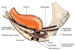
- Layered musculotendinous structure between the Anal Canal and the caudal part of the vertebral column, on which the terminal Rectum sits
- From above downwards, it consists of:
- Superior Fascia of the Pelvic Diaphragm
- Tendinous Plate of Pubococcygeus
- Muscular Raphe of Iliococcygeus
- Posterior parts of Puborectalis and the External Anal Sphincter
- Nerve Supply:
- Main supply: Sacral Plexus by Branches of S3 and S4
- Enter the upper (pelvic) surface of the muscle
- Some fibres travel in or close to Pelvic Splanchnic Nerves
- Puborectalis, Pubourethralis, Pubovaginalis supplied from below by the Perineal Branch of S4 and Inferior Rectal Branch of Pudendal Nerve, in common with the External Anal Sphincter
- High proportion of slow twitch fibres (like the External Anal Sphincter and Urethral Sphincter muscles)
- Main supply: Sacral Plexus by Branches of S3 and S4
Coccygeus
- Best thought of as the Ischiococcygeus
- Origin: Tip of the Ischial Spine
- Insertion: Side of Coccyx and lowest piece of Sacrum
- Lies edge to edge with the lower border of Piriformis
- Overlapped anteriorly by Iliococcygeus
- Gluteal surface is fibrous tissue – is indeed the Sacrospinous Ligament
- Nerve supply:
- Branches of S3 and S4
Perineal Body
-
a.k.a. Central Tendon of the Perineum
-
Midline fibromuscular mass to which a number of muscles gain attachment and within which they decussate
-
Attached to the posterior border of the Perineal Membrane
-
Lies between the Anal Canal and the Vagina or the Bulb of the Penis
-
Rectovaginal Septum blends into it above
-
Muscles running into it include:
- External Anal Sphincter
- Pubovaginalis/ Puboprostaticus part of Levator Ani
- Bulbospongiosus
- Superficial and Deep Transverse Perineal Muscles
-
Position and connections provide stabilizing influence for pelvic and perineal structures
-
Injury at childbirth may weak pelvic floor contributing to prolapse of Vagina and Uterus
Pelvic fascia
Parietal Pelvic Fascia
- On pelvic surface of Obturator Internus
- Strong membrane
- Fuses with the Periosteum at the upper margin of the Obturator Internus muscle
- Becomes thin below the Tendinous Arch where it covers Obturator Internus on the lateral wall of the Ischioanal Fossa
- Fascia on pelvic surface of Piriformis fuses with the periosteum at the medial margins of the Anterior Sacral Foramina
- Sacral Anterior Primary Rami emerges from these foramina thus lie behind the fascia
- Internal Iliac Vessels lie in front of the fascia over Piriformis
- Large Pre-sacral Lateral Sacral Veins lie initially behind this fascia as they emerge from the Anterior Sacral Foramina
Rectosacral Fascia
-
A condensation of connective tissue from the front of the lower Sacrum
-
Passes downwards and forwards to fuse with the Mesorectal Fascia 3-5cm proximal to the Anorectal Junction
-
Large Pre-Sacral Lateral Sacral Veins lie behind this fascia on the front of the Sacrum
-
Superior Fascia of the Pelvic Diaphragm
- Fascia on the pelvic surface of Levator Ani and Coccygeus
- Attached in front to the posterior surface of the Body of the Pubis and at the back to the Ischial Spine
- In between attachments, it blends with the Obturator Fascia
- Thickening of these two fused fasciae forms the Tendinous Arch of Origin of Levator Ani
-
Inferior Fascia of the Pelvic Diaphragm
- Thin fascia covering the undersurface of Levator Ani
- On the sloping medial wall of the Ischioanal Fossa
- Blends with the Obturator Fascia laterally
- Blends with the fascia on the External Anal and Urethral Sphincters medially
Sensation
- Pudendal nerve (S2–S4) – primary somatic nerve of the perineum
- Exits pelvis via greater sciatic foramen, curves around ischial spine, re-enters via lesser sciatic foramen
- Travels through pudendal (Alcock’s) canal
- Branches:
- Inferior rectal nerve – to anal canal and external anal sphincter
- Perineal nerve – to perineal muscles, posterior scrotum/labia, skin of perineal body
- Dorsal nerve of penis/clitoris – sensory to glans, body of penis/clitoris - Ilioinguinal nerve (L1)
- Supplies skin of the root of the penis or mons pubis, and anterior scrotum/labia majora - Genitofemoral nerve (L1–L2)
- Genital branch supplies the cremaster muscle and skin of anterior scrotum/labia majora - Posterior femoral cutaneous nerve (S1–S3)
- Perineal branch (aka inferior cluneal) supplies posterior scrotum/labia and inferior gluteal fold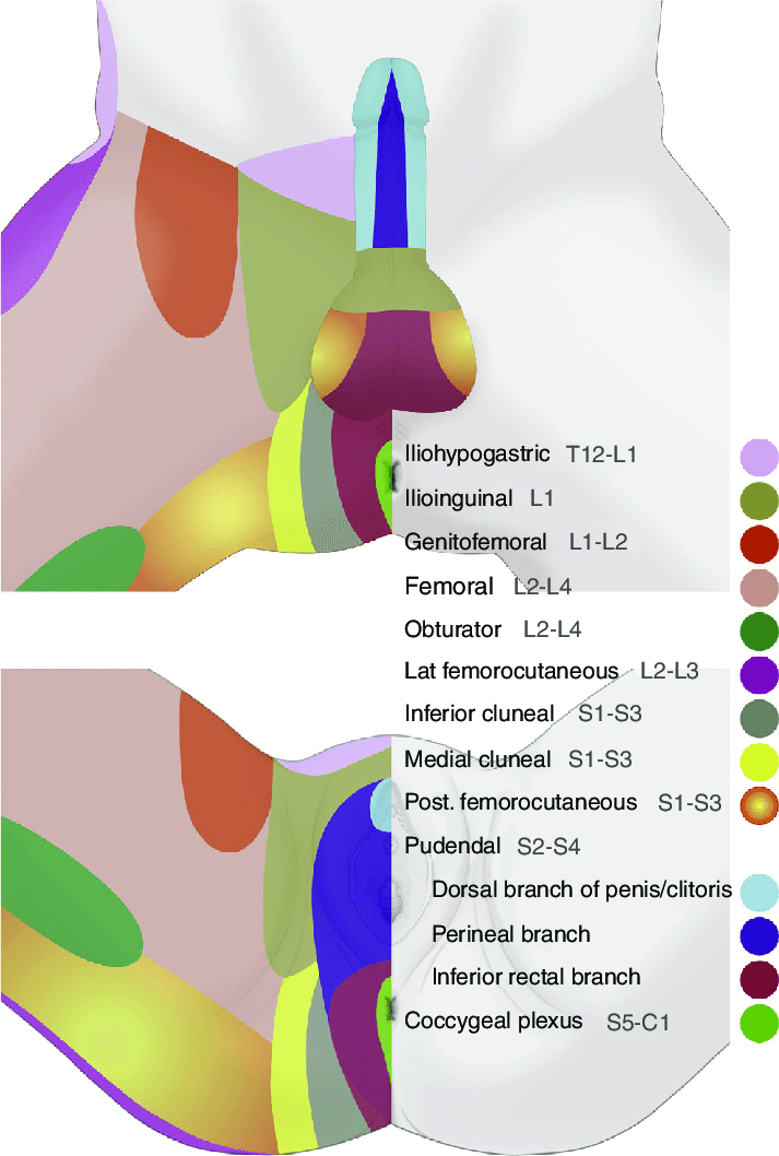
Blood Vessels
Internal Iliac Artery
-
Origin: Common Iliac Artery which bifurcates at pelvic brim opposite Sacroiliac joint
-
Divides into short Posterior and longer Anterior division
-
Posterior division
-
Anterior division
- Bladder
- Superior Vesical Artery +/- Obliterated Umbilical Artery
- Inferior Vesical Artery (Some textbook says only in males)
- Other visceral
- Parietal
- Bladder