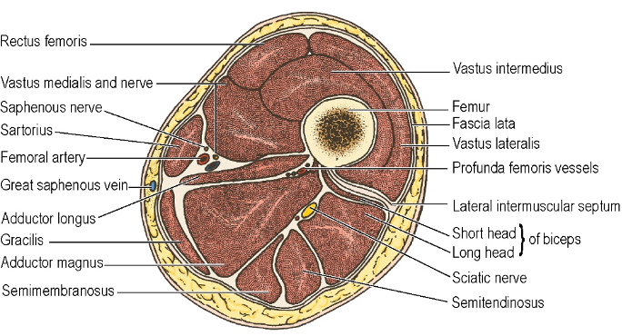
Femoral canal anatomy
Boundaries
- Proximal opening: Femoral Ring
- Boundaries of Femoral Ring:
- Anterior: Medial part of Inguinal Ligament
- Medial: Crescentic edge of Lacunar Ligament
- Posterior: Pectineal Ligament
- Lateral: Femoral Vein
- Boundaries of Femoral Ring:
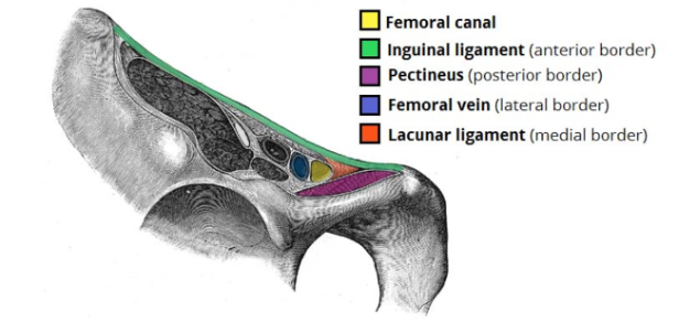
- 25% of pts have an Aberrant Obturator Artery (Corona Mortis) which can cause significant bleeding if the Lacunar Ligament is stretched or divided during femoral hernia repair
- Aberrant Obturator Artery a branch of the Inferior Epigastric Artery
Contents
-
Lymph node (of Cloquet)
- Female: Drains directly from Clitoris
- Male: Drains from the Glans
-
Lymphatic vessels draining Deep Inguinal Lymph Nodes
-
Empty space
-
Loose connective tissue
-
NB: In Femoral Hernias, the Hernial Sac:
- Passes behind the Inguinal Ligament
- Passes medial to the Femoral Vein
- Passes anterior to the Superior Pubic Ramus
- Covered by the fascias of the Femoral Septum and Cribriform Fascia
- Contains a large quantity of fat
- More common in female than in male patients
- Less common than inguinal hernias in female patients
- Not able to be controlled by wearing a hernia truss
- Classically associated with strangulation bowel obstruction of Richter type
- Not more prone to recurrence after surgery than inguinal hernias
Femoral triangle
Boundaries
- Lateral: Sartorius (medial border)
- Medial: Adductor Longus (medial border)
- Superior: Inguinal Ligament
- Floor: Iliacus, Psoas, Pectineus and Adductor Longus
- NB: Roof: Fascia Lata of the Thigh
Contents
- Femoral Nerve
- Femoral Artery
- Femoral Vein
- Femoral Canal containing lymph nodes and lymphatics and loose connective tissue
Femoral Sheath
- Funnel shaped prolongation of the extraperitoneal fascia surrounding the femoral vessels
- Passing beneath the inguinal ligament
- Formed by
- Transversalis Fascia anteriorly
- Psoas fascia posteriorly
- Both fuse with adventitia of Femoral Artery and Vein, one inch below inguinal ligament
- Sheath allows freedom of movement of these vessels
- Has three compartments
- Lateral (Femoral Artery and Femoral branch Genitofemoral Nerve)
- Intermediate (Femoral Vein)
- Medial (Femoral Canal)
- NB: Femoral nerve is not within the Femoral Sheath
- It’s lateral
Femoral Artery
Course
- Enters thigh at point midway between Anterior Superior Iliac Spine (ASIS) and Pubic Symphysis (Mid-Inguinal point)
- Lies on Psoas Major tendon, which separates it from capsule of hip joint
- This is the point where its pulse can be felt and where arterial catheterization can occur
- Emerges from Femoral Sheath
- Courses downwards and enters Adductor Canal deep to Sartorius
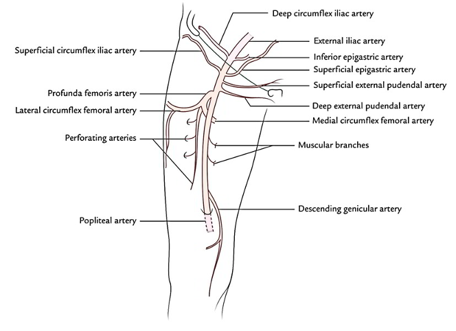
Branches
- Four small branches below Inguinal Ligament
- One large branch just below the termination of the Femoral Sheath
- Profunda Femoris – chief artery of the thigh
Surface Marking
- With the hip slightly flexed and laterally rotated
- Midpoint between Pubic Symphysis and ASIS along the upper 2/3rd of a line towards the Adductor Tubercle
Surgical Approach
- In the Adductor Canal through an incision on the medial aspect of the lower thigh
- Sartorius retracted medially and fascial roof of canal divided
Femoral Vein
- Enters lower angle of Femoral Triangle (where it lies posterior to artery)
- Ascends through Femoral triangle
- Comes to lie on medial side of Femoral Artery
- Receives Profunda Femoris vein posteriorly
- Receives Great Saphenous Vein anteriorly just below the Femoral Sheath
- Within the Femoral Sheath, it passes under the Inguinal Ligament
- Runs along the pelvic brim as the External Iliac Vein
Femoral nerve
Popliteal Artery
- Throughout course, it is the deepest neurovascular structure in the Popliteal Fossa
- Extends from Adductor Hiatus in Adductor Magnus (a hand’s breadth above the knee) to the fibrous arch of Soleus (a hand’s breadth below the knee)
- Enters Popliteal Fossa on medial side of Femur
- Here, it lies deep and medial to Sciatic Nerve and its vertical continuation, the Tibial Nerve
- Convexes laterally as it passes downwards
- Comes to lie lateral to the Tibial Nerve
- Below fibrous arch of Soleus, (as the Posterior Tibial Artery) it returns to the medial side of the Tibial Nerve
- Below fibrous arch of Soleus, it immediately divides into Anterior tibial artery and Posterior tibial artery
- Artery lies successively on popliteal surface of Femur (separated from it by a little fat), on the Oblique Popliteal Ligament and on the fascia of Popliteus
- Palpation: Centre of the popliteal fossa (with the knee slightly flexed to relax the popliteal fascia)
Popliteal Fossa
- Diamond-shaped space behind the knee
- Boundaries:
- Superior Medial: Semimembranosus, Semitendinosus
- Superior Lateral: Biceps Femoris
- Inferior: Heads of Gastrocnemius
- Roof: Fascia Lata
- Roof pierced by: Small Saphenous Vein and Posterior Femoral Cutaneous Nerve
- Floor (from above downwards)
- Femur (popliteal surface)
- Capsule of Knee Joint
- Oblique Popliteal Ligament
- Popliteus
- Small group of Popliteal Lymph Nodes lie alongside the Popliteal Vein
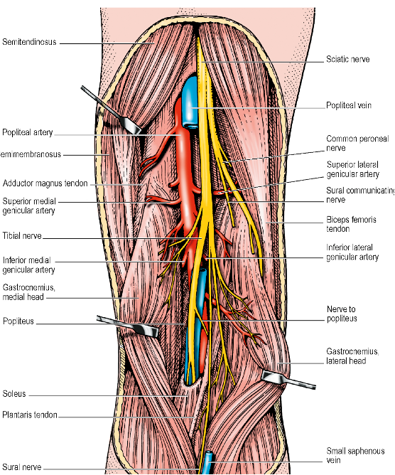
Contents (medial to lateral)
Adductor Canal
- a.k.a. Subsartorial or Hunter’s Canal
- Extending from the apex of the femoral triangle to the Adductor Hiatus of the adductor magnus.
- Groove between Vastus Medialis and front of Adductor muscles below the apex of the Femoral Triangle
- Lateral: Vastus Medialis
- Anteriormedial: Sartorius
- Floor: Adductor Longus (above) and Adductor Magnus (below)
- Contents:
- Femoral Artery
- Femoral Vein
- Saphenous Nerve
- Nerve to Vastus Medialis